2P5N image
Deposition Date 2007-03-15
Release Date 2007-10-09
Last Version Date 2024-11-13
Entry Detail
PDB ID:
2P5N
Keywords:
Title:
Crystal structure of mouse 17-alpha hydroxysteroid dehydrogenase in complex with coenzyme NADPH
Biological Source:
Source Organism:
Mus musculus (Taxon ID: 10090)
Host Organism:
Method Details:
Experimental Method:
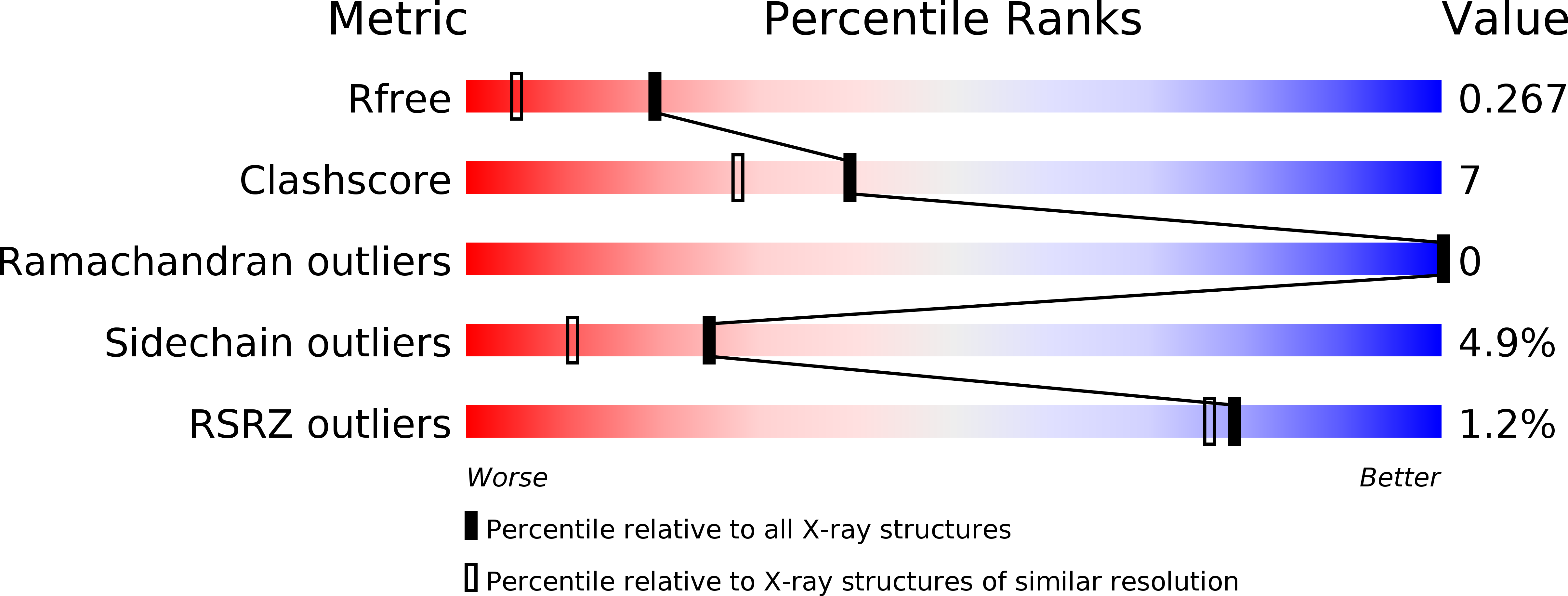
Resolution:
1.80 Å
R-Value Free:
0.26
R-Value Work:
0.19
R-Value Observed:
0.20
Space Group:
P 21 21 21
Macromolecular Entities
Structures with similar UniProt ID
Protein Blast
Polymer Type:polypeptide(L)
Molecule:Aldo-keto reductase family 1, member C21
Chain IDs:A, B
Chain Length:323
Number of Molecules:2
Biological Source:Mus musculus
Primary Citation
Structure of 3(17)alpha-hydroxysteroid dehydrogenase (AKR1C21) holoenzyme from an orthorhombic crystal form: an insight into the bifunctionality of the enzyme.
Acta Crystallogr.,Sect.F 63 825 830 (2007)
PMID: 17909281 DOI: 10.1107/S1744309107040985

Abstact

Mouse 3(17)alpha-hydroxysteroid dehydrogenase (AKR1C21) is a bifunctional enzyme that catalyses the oxidoreduction of the 3- and 17-hydroxy/keto groups of steroid substrates such as oestrogens, androgens and neurosteroids. The structure of the AKR1C21-NADPH binary complex was determined from an orthorhombic crystal belonging to space group P2(1)2(1)2(1) at a resolution of 1.8 A. In order to identify the factors responsible for the bifunctionality of AKR1C21, three steroid substrates including a 17-keto steroid, a 3-keto steroid and a 3alpha-hydroxysteroid were docked into the substrate-binding cavity. Models of the enzyme-coenzyme-substrate complexes suggest that Lys31, Gly225 and Gly226 are important for ligand recognition and orientation in the active site.

Legend

Protein

Chemical

Disease

Primary Citation of related structures
Feedback Form
Name
Email
Institute
Feedback