2P4N image
Deposition Date 2007-03-12
Release Date 2008-07-08
Last Version Date 2024-02-21
Entry Detail
PDB ID:
2P4N
Title:
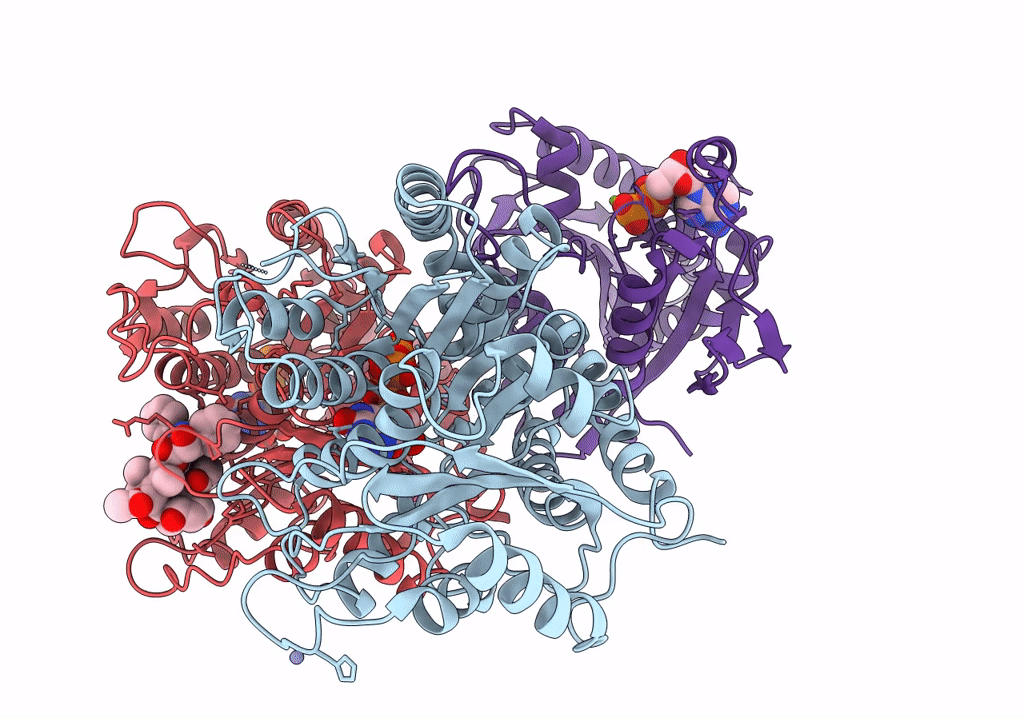
Human Monomeric Kinesin (1BG2) and Bovine Tubulin (1JFF) Docked into the 9-Angstrom Cryo-EM Map of Nucleotide-Free Kinesin Complexed to the Microtubule
Biological Source:
Source Organism:
Homo sapiens (Taxon ID: )
Method Details:
Experimental Method:
Resolution:
9.00 Å
Aggregation State:
FILAMENT
Reconstruction Method:
SINGLE PARTICLE
Macromolecular Entities
Polymer Type:polypeptide(L)
Molecule:Tubulin alpha chain
Chain IDs:B (auth: A)
Chain Length:451
Number of Molecules:1
Biological Source:Bos taurus
Polymer Type:polypeptide(L)
Molecule:Tubulin beta chain
Chain IDs:C (auth: B)
Chain Length:445
Number of Molecules:1
Biological Source:Bos taurus
Polymer Type:polypeptide(L)
Molecule:Kinesin heavy chain
Chain IDs:A (auth: K)
Chain Length:325
Number of Molecules:1
Biological Source:Homo sapiens
Primary Citation
The beginning of kinesin's force-generating cycle visualized at 9-A resolution.
J.Cell Biol. 177 377 385 (2007)
PMID: 17470637 DOI: 10.1083/jcb.200612090

Abstact

We have used cryo-electron microscopy of kinesin-decorated microtubules to resolve the structure of the motor protein kinesin's crucial nucleotide response elements, switch I and the switch II helix, in kinesin's poorly understood nucleotide-free state. Both of the switch elements undergo conformational change relative to the microtubule-free state. The changes in switch I suggest a role for it in "ejecting" adenosine diphosphate when kinesin initially binds to the microtubule. The switch II helix has an N-terminal extension, apparently stabilized by conserved microtubule contacts, implying a microtubule activation mechanism that could convey the state of the bound nucleotide to kinesin's putative force-delivering element (the "neck linker"). In deriving this structure, we have adapted an image-processing technique, single-particle reconstruction, for analyzing decorated microtubules. The resulting reconstruction visualizes the asymmetric seam present in native, 13-protofilament microtubules, and this method will provide an avenue to higher-resolution characterization of a variety of microtubule- binding proteins, as well as the microtubule itself.

Legend

Protein

Chemical

Disease

Primary Citation of related structures