Deposition Date
2007-03-12
Release Date
2007-05-22
Last Version Date
2024-03-13
Entry Detail
Biological Source:
Source Organism(s):
Escherichia coli K12 (Taxon ID: 83333)
Expression System(s):
Method Details:
Experimental Method:
Resolution:
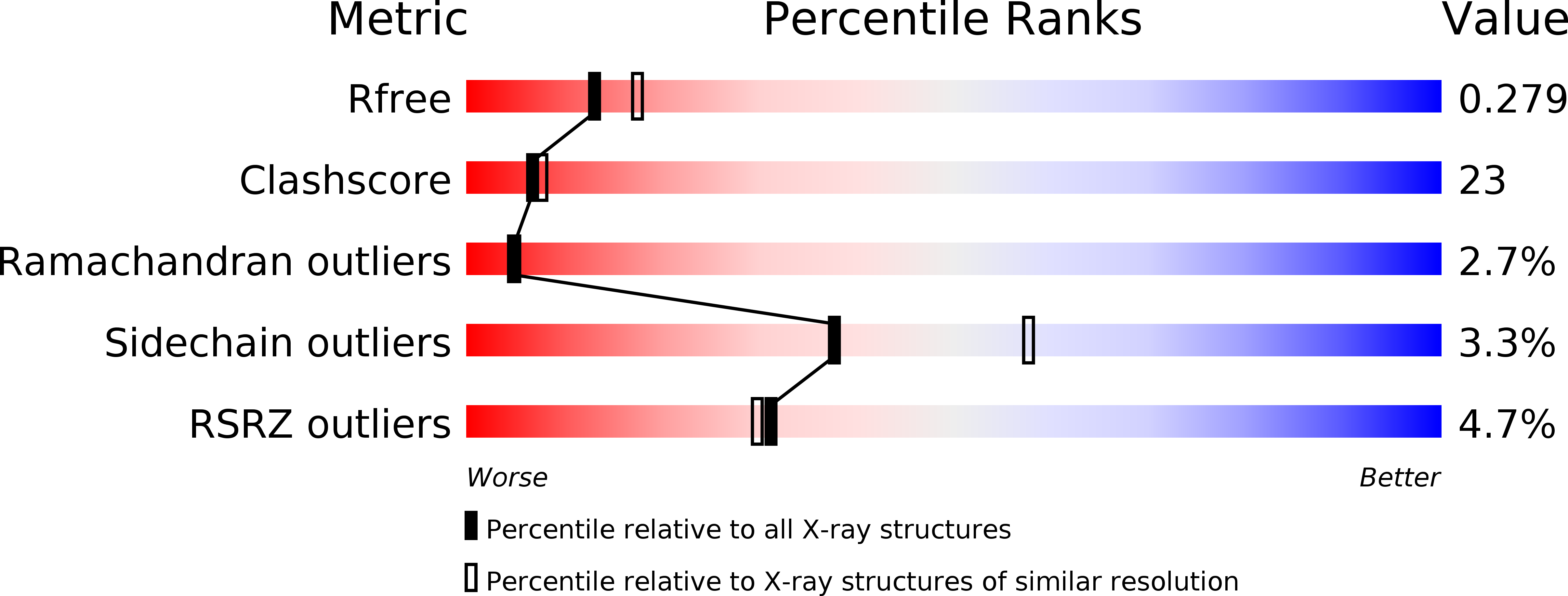
2.40 Å
R-Value Free:
0.28
R-Value Work:
0.23
R-Value Observed:
0.23
Space Group:
C 2 2 21