2P1W image
Entry Detail
PDB ID:
2P1W
Keywords:
Title:
structure of the phosphothreonine lyase SpvC, the effector protein from Salmonella
Biological Source:
Source Organism:
PDB Version:
Deposition Date:
2007-03-06
Release Date:
2007-12-11
Method Details:
Experimental Method:
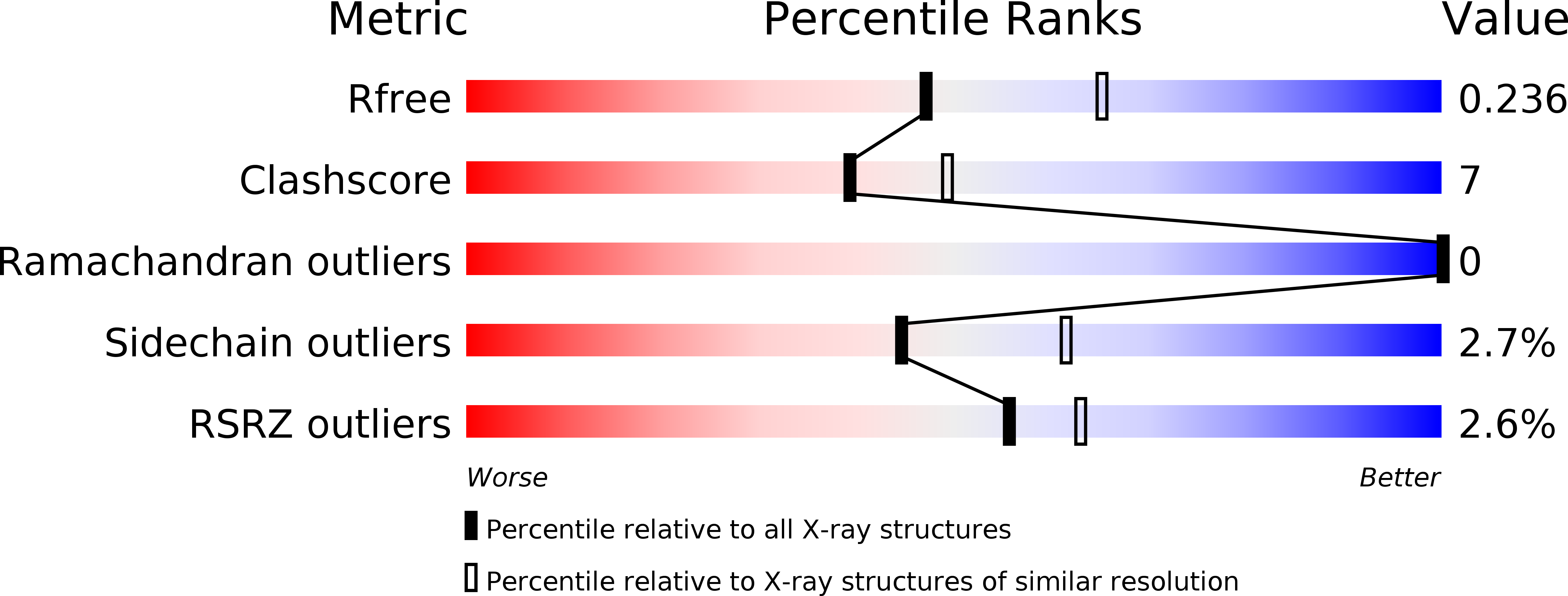
Resolution:
2.30 Å
R-Value Free:
0.22
R-Value Work:
0.19
Space Group:
P 42 21 2
Macromolecular Entities
Polymer Type:polypeptide(L)
Description:27.5 kDa virulence protein
Chain IDs:A
Chain Length:250
Number of Molecules:1
Biological Source:Salmonella enteritidis
Modified Residue
Compound ID Chain ID Parent Comp ID Details 2D Image
MSE A MET SELENOMETHIONINE
Primary Citation
Structural insights into the enzymatic mechanism of the pathogenic MAPK phosphothreonine lyase.
Mol.Cell 28 899 913 (2007)
PMID: 18060821 DOI: 10.1016/j.molcel.2007.11.011

Abstact

The OspF family of phosphothreonine lyase, including SpvC from Salmonella, irreversibly inactivates the dual-phosphorylated host MAPKs (pT-X-pY) through beta elimination. We determined crystal structures of SpvC and its complex with a phosphopeptide substrate. SpvC adopts a unique fold of alpha/beta type. The disordered N terminus harbors a canonical D motif for MAPK substrate docking. The enzyme-substrate complex structure indicates that recognition of the phosphotyrosine followed by insertion of the threonine phosphate into an arginine pocket places the phosphothreonine into the enzyme active site. This requires the conformational flexibility of pT-X-pY, which suggests that p38 (pT-G-pY) is likely the preferred physiological substrate. Structure-based biochemical and enzymatic analysis allows us to propose a general acid/base mechanism for beta elimination reaction catalyzed by the phosphothreonine lyase. The mechanism described here provides a structural understanding of MAPK inactivation by a family of pathogenic effectors conserved in plant and animal systems and may also open a new route for biological catalysis.

Legend

Protein

Chemical

Disease

Primary Citation of related structures