2OWL image
Deposition Date 2007-02-16
Release Date 2007-03-20
Last Version Date 2024-10-09
Entry Detail
PDB ID:
2OWL
Keywords:
Title:
Crystal structure of E. coli RdgC
Biological Source:
Source Organism:
Escherichia coli (Taxon ID: 562)
Host Organism:
Method Details:
Experimental Method:
Resolution:
2.40 Å
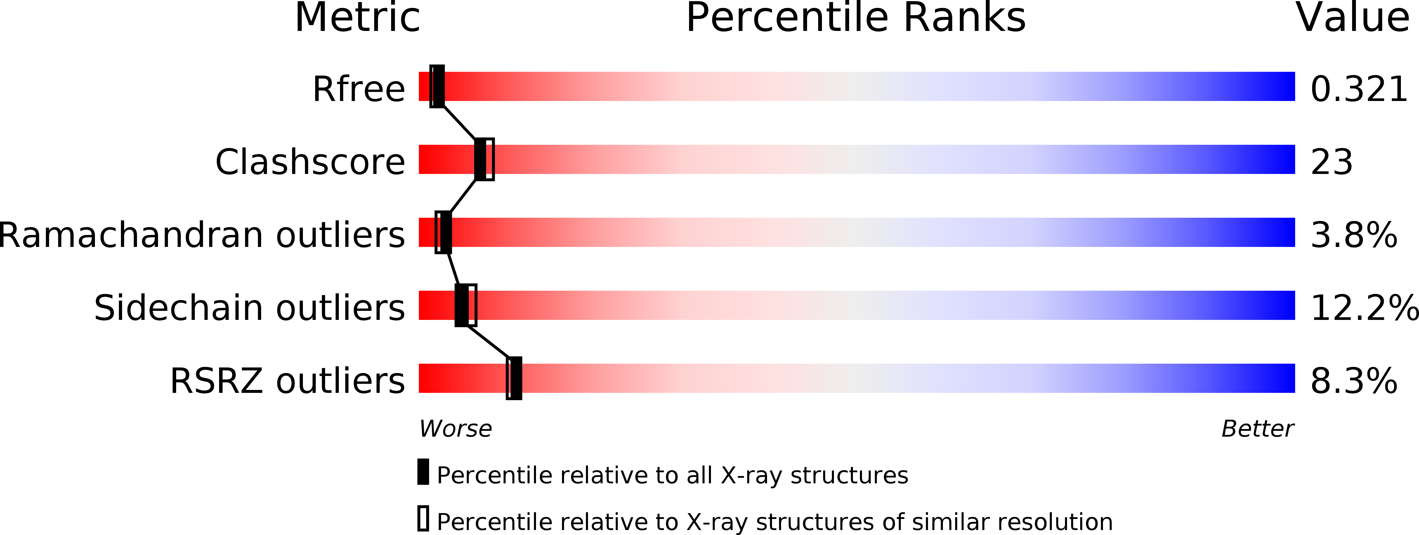
R-Value Free:
0.32
R-Value Work:
0.23
R-Value Observed:
0.24
Space Group:
P 21 21 21
Macromolecular Entities
Structures with similar UniProt ID
Protein Blast
Polymer Type:polypeptide(L)
Molecule:Recombination-associated protein rdgC
Gene (Uniprot):rdgC
Chain IDs:A, B
Chain Length:303
Number of Molecules:2
Biological Source:Escherichia coli
Modified Residue
Compound ID Chain ID Parent Comp ID Details 2D Image
MSE A MET SELENOMETHIONINE
Ligand Molecules
Primary Citation
Ring Structure of the Escherichia coli DNA-binding Protein RdgC Associated with Recombination and Replication Fork Repair.
J.Biol.Chem. 282 12353 12357 (2007)
PMID: 17308310 DOI: 10.1074/jbc.C700023200

Abstact

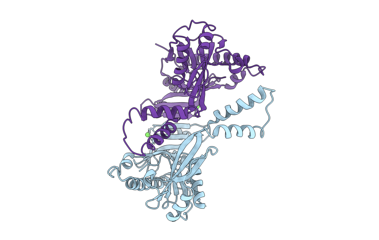
The DNA-binding protein, RdgC, is associated with recombination and replication fork repair in Escherichia coli and with the virulence-associated, pilin antigenic variation mediated by RecA and other recombination proteins in Neisseria species. We solved the structure of the E. coli protein and refined it to 2.4A. RdgC crystallizes as a dimer with a head-to-head, tail-to-tail organization forming a ring with a 30 A diameter hole at the center. The protein fold is unique and reminiscent of a horseshoe with twin gates closing the open end. The central hole is lined with positively charged residues and provides a highly plausible DNA binding channel consistent with the nonspecific mode of binding detected in vitro and with the ability of RdgC to modulate RecA function in vivo.

Legend

Protein

Chemical

Disease

Primary Citation of related structures