
Deposition Date
2007-02-16
Release Date
2007-06-19
Last Version Date
2024-02-21
Entry Detail
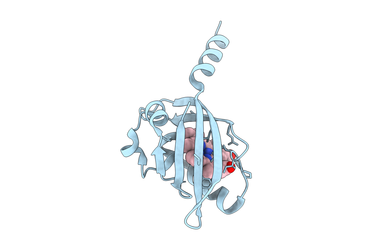
PDB ID:
2OWH
Keywords:
Title:
Structure of an early-microsecond photolyzed state of CO-bjFixLH
Biological Source:
Source Organism(s):
Bradyrhizobium japonicum (Taxon ID: 375)
Expression System(s):
Method Details:
Experimental Method:
Resolution:
2.50 Å
R-Value Free:
0.30
R-Value Work:
0.23
R-Value Observed:
0.24
Space Group:
H 3 2
