
Deposition Date
2007-02-15
Release Date
2007-04-24
Last Version Date
2024-02-21
Entry Detail
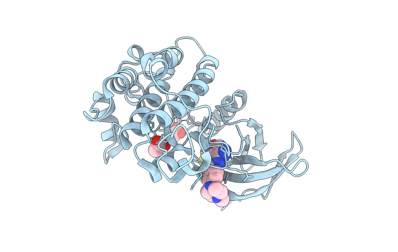
PDB ID:
2OWB
Keywords:
Title:
Structure of the Catalytic Domain of Human Polo-like Kinase 1
Biological Source:
Source Organism(s):
Homo sapiens (Taxon ID: 9606)
Expression System(s):
Method Details:
Experimental Method:
Resolution:
2.10 Å
R-Value Free:
0.24
R-Value Work:
0.20
R-Value Observed:
0.20
Space Group:
P 32 2 1
