Deposition Date
2007-02-15
Release Date
2008-01-08
Last Version Date
2024-11-20
Entry Detail
PDB ID:
2OW6
Keywords:
Title:
Golgi alpha-mannosidase II complex with (1r,5s,6s,7r,8s)-1-thioniabicyclo[4.3.0]nonan-5,7,8-triol chloride
Biological Source:
Source Organism(s):
Drosophila melanogaster (Taxon ID: 7227)
Expression System(s):
Method Details:
Experimental Method:
Resolution:
1.19 Å
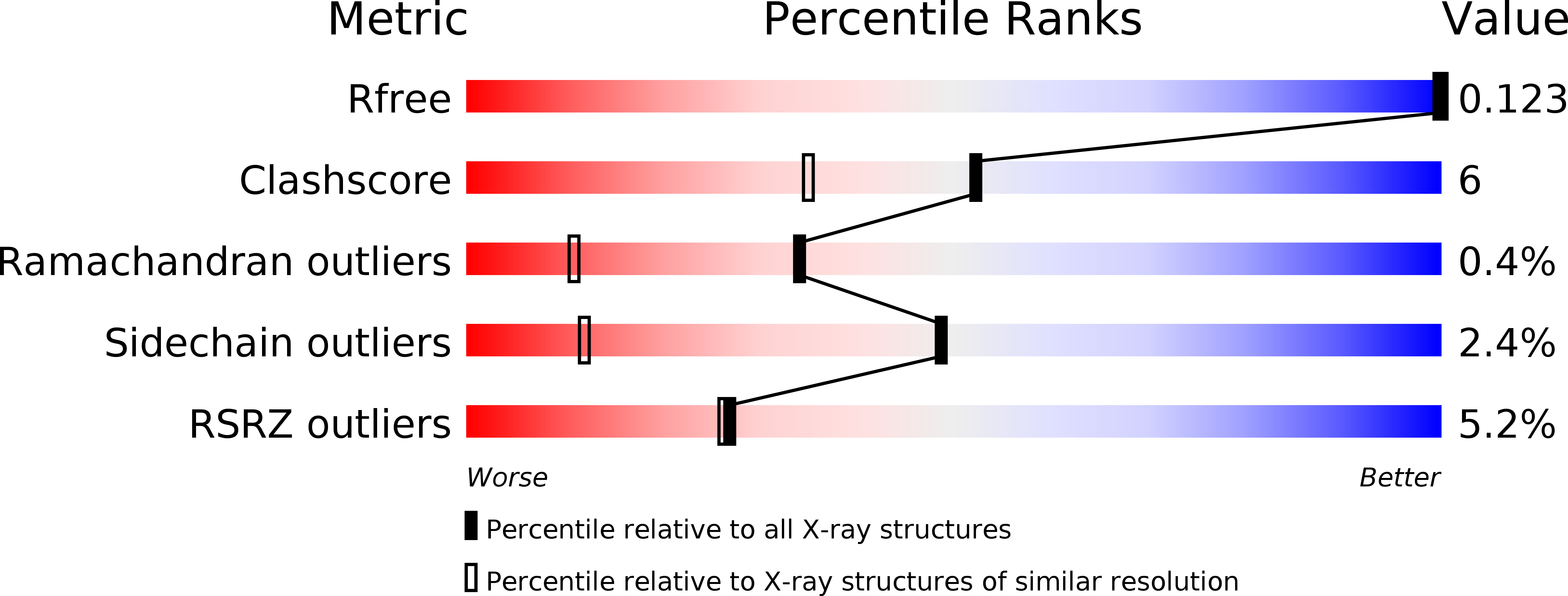
R-Value Free:
0.15
R-Value Work:
0.11
R-Value Observed:
0.11
Space Group:
P 21 21 21