2OVI image
Deposition Date 2007-02-13
Release Date 2008-02-12
Last Version Date 2024-02-21
Entry Detail
PDB ID:
2OVI
Title:
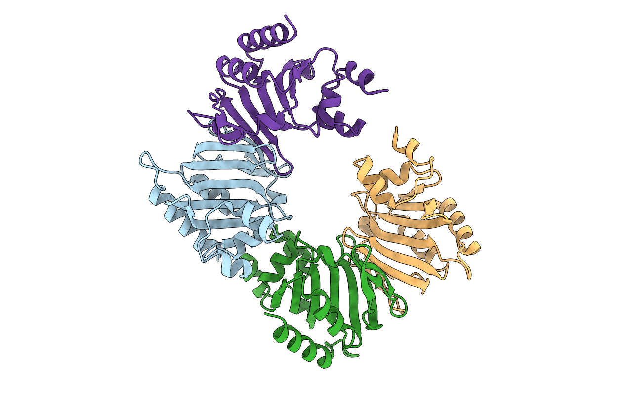
Structure of the Heme Binding Protein ChuX
Biological Source:
Source Organism:
Method Details:
Experimental Method:
Resolution:
2.05 Å
R-Value Free:
0.25
R-Value Work:
0.21
Space Group:
P 41
Macromolecular Entities
Structures with similar UniProt ID
Protein Blast
Polymer Type:polypeptide(L)
Molecule:Hypothetical protein chuX
Gene (Uniprot):ECs_4384
Chain IDs:A, B, C, D
Chain Length:164
Number of Molecules:4
Biological Source:Escherichia coli O157:H7
Primary Citation
Structure and heme binding properties of Escherichia coli O157:H7 ChuX.
Protein Sci. 18 825 838 (2009)
PMID: 19319934 DOI: 10.1002/pro.84

Abstact

For many pathogenic microorganisms, iron acquisition from host heme sources stimulates growth, multiplication, ultimately enabling successful survival and colonization. In gram-negative Escherichia coli O157:H7, Shigella dysenteriae and Yersinia enterocolitica the genes encoded within the heme utilization operon enable the effective uptake and utilization of heme as an iron source. While the complement of proteins responsible for heme internalization has been determined in these organisms, the fate of heme once it has reached the cytoplasm has only recently begun to be resolved. Here we report the first crystal structure of ChuX, a member of the conserved heme utilization operon from pathogenic E. coli O157:H7 determined at 2.05 A resolution. ChuX forms a dimer which remarkably given low sequence homology, displays a very similar fold to the monomer structure of ChuS and HemS, two other heme utilization proteins. Absorption spectral analysis of heme reconstituted ChuX demonstrates that ChuX binds heme in a 1:1 manner implying that each ChuX homodimer has the potential to coordinate two heme molecules in contrast to ChuS and HemS where only one heme molecule is bound. Resonance Raman spectroscopy indicates that the heme of ferric ChuX is composed of a mixture of coordination states: 5-coordinate and high-spin, 6-coordinate and low-spin, and 6-coordinate and high-spin. In contrast, the reduced ferrous form displays mainly a 5-coordinate and high-spin state with a minor contribution from a 6-coordinate and low-spin state. The nu(Fe-CO) and nu(C-O) frequencies of ChuX-bound CO fall on the correlation line expected for histidine-coordinated hemoproteins indicating that the fifth axial ligand of the ferrous heme is the imidazole ring of a histidine residue. Based on sequence and structural comparisons, we designed a number of site-directed mutations in ChuX to probe the heme binding sites and dimer interface. Spectral analysis of ChuX and mutants suggests involvement of H65 and H98 in heme coordination as mutations of both residues were required to abolish the formation of the hexacoordination state of heme-bound ChuX.

Legend

Protein

Chemical

Disease

Primary Citation of related structures