Deposition Date
2007-02-13
Release Date
2007-02-27
Last Version Date
2023-08-30
Entry Detail
PDB ID:
2OVB
Keywords:
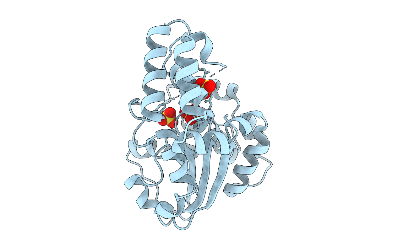
Title:
Crystal Structure of StaL-sulfate complex
Biological Source:
Source Organism(s):
Streptomyces toyocaensis (Taxon ID: 55952)
Expression System(s):
Method Details:
Experimental Method:
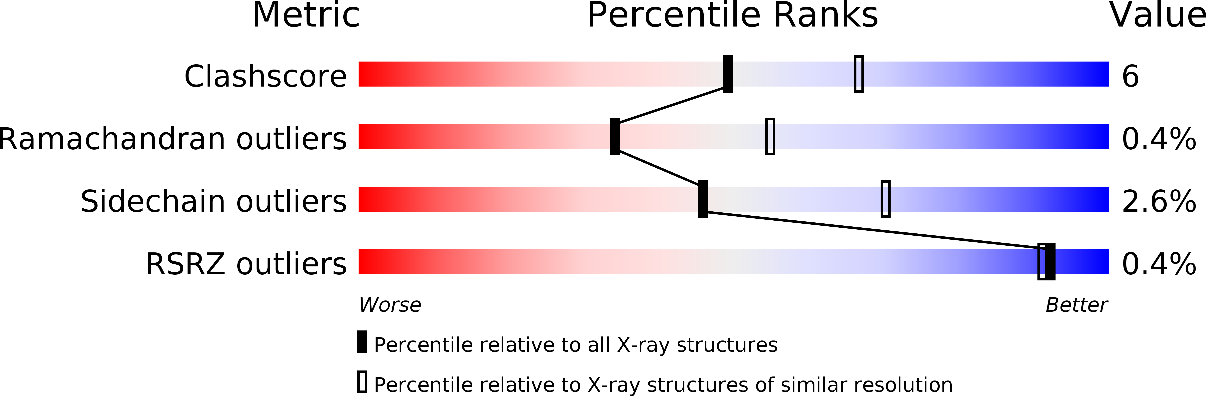
Resolution:
2.61 Å
R-Value Free:
0.26
R-Value Work:
0.22
R-Value Observed:
0.23
Space Group:
P 61 2 2