Deposition Date
2007-02-10
Release Date
2007-05-01
Last Version Date
2024-02-21
Entry Detail
PDB ID:
2OUG
Keywords:
Title:
Crystal structure of the RfaH transcription factor at 2.1A resolution
Biological Source:
Source Organism(s):
Escherichia coli (Taxon ID: 562)
Expression System(s):
Method Details:
Experimental Method:
Resolution:
2.10 Å
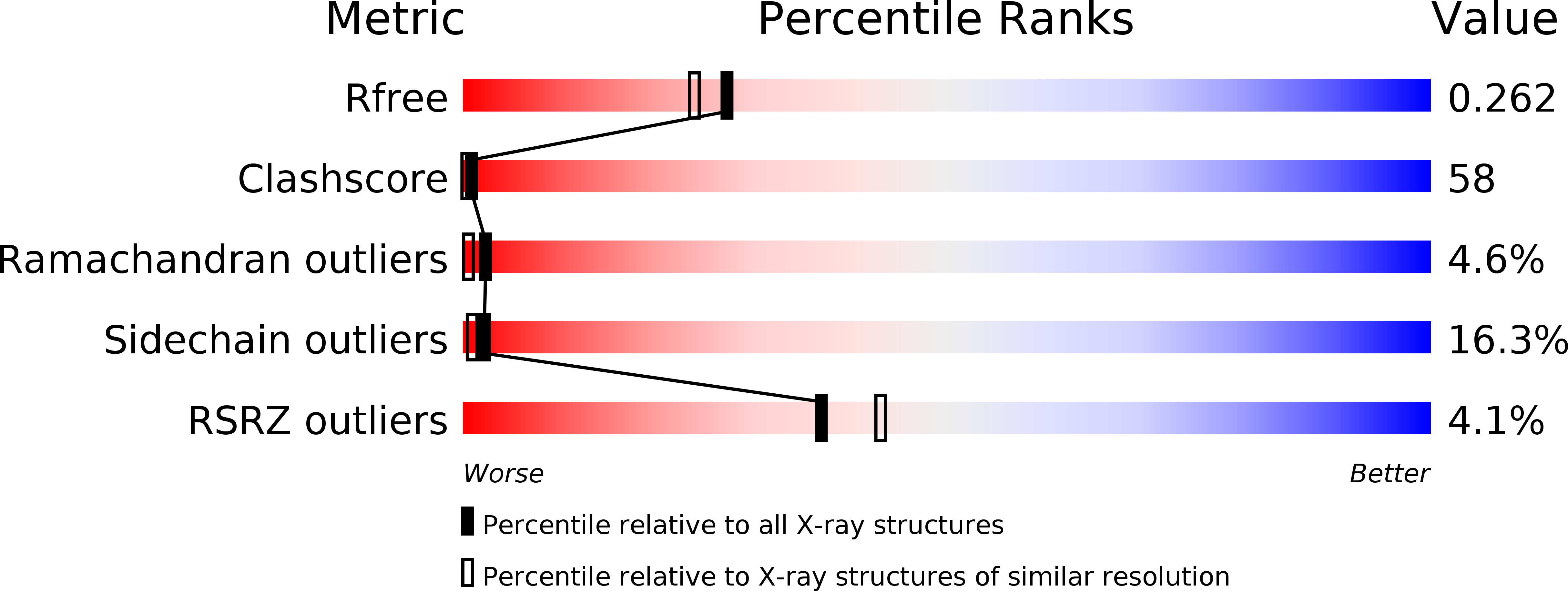
R-Value Free:
0.27
R-Value Work:
0.23
R-Value Observed:
0.24
Space Group:
P 65