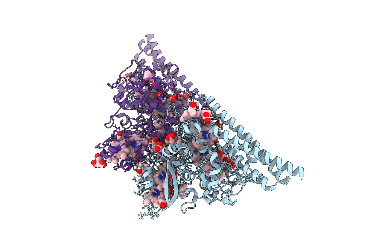
2OT4 image
Deposition Date 2007-02-07
Release Date 2008-04-08
Last Version Date 2024-11-20
Entry Detail
PDB ID:
2OT4
Keywords:
Title:
Structure of a hexameric multiheme c nitrite reductase from the extremophile bacterium Thiolkalivibrio nitratireducens
Biological Source:
Source Organism:
Method Details:
Experimental Method:
Resolution:
1.50 Å
R-Value Free:
0.14
R-Value Work:
0.12
R-Value Observed:
0.12
Space Group:
P 21 3
Macromolecular Entities
Polymer Type:polypeptide(L)
Molecule:Eight-heme nitrite reductase
Chain IDs:A, B
Chain Length:525
Number of Molecules:2
Biological Source:Thioalkalivibrio nitratireducens
Primary Citation

Abstact

Bacterial pentaheme cytochrome c nitrite reductases (NrfAs) are key enzymes involved in the terminal step of dissimilatory nitrite reduction of the nitrogen cycle. Their structure and functions are well studied. Recently, a novel octaheme cytochrome c nitrite reductase (TvNiR) has been isolated from the haloalkaliphilic bacterium Thioalkalivibrio nitratireducens. Here we present high-resolution crystal structures of the apoenzyme and its complexes with the substrate (nitrite) and the inhibitor (azide). Both in the crystalline state and in solution, TvNiR exists as a stable hexamer containing 48 hemes-the largest number of hemes accommodated within one protein molecule known to date. The subunit of TvNiR consists of two domains. The N-terminal domain has a unique fold and contains three hemes. The catalytic C-terminal domain hosts the remaining five hemes, their arrangement, including the catalytic heme, being identical to that found in NrfAs. The complete set of eight hemes forms a spatial pattern characteristic of other multiheme proteins, including structurally characterized octaheme cytochromes. The catalytic machinery of TvNiR resembles that of NrfAs. It comprises the lysine residue at the proximal position of the catalytic heme, the catalytic triad of tyrosine, histidine, and arginine at the distal side, channels for the substrate and product transport with a characteristic gradient of electrostatic potential, and, finally, two conserved Ca(2+)-binding sites. However, TvNiR has a number of special structural features, including a covalent bond between the catalytic tyrosine and the adjacent cysteine and the unusual topography of the product channels that open into the void interior space of the protein hexamer. The role of these characteristic structural features in the catalysis by this enzyme is discussed.

Legend

Protein

Chemical

Disease

Primary Citation of related structures