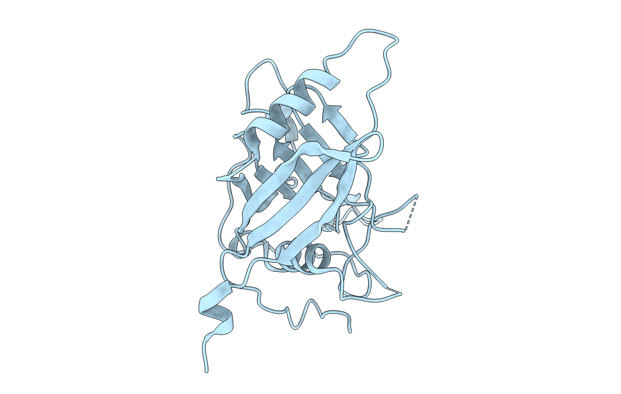
2OSE image
Deposition Date 2007-02-05
Release Date 2007-12-18
Last Version Date 2023-08-30
Entry Detail
PDB ID:
2OSE
Keywords:
Title:
Crystal Structure of the Mimivirus Cyclophilin
Biological Source:
Source Organism:
Mimivirus (Taxon ID: 315393)
Host Organism:
Method Details:
Experimental Method:
Resolution:
2.04 Å
R-Value Free:
0.25
R-Value Work:
0.21
R-Value Observed:
0.21
Space Group:
P 21 3
Macromolecular Entities
Structures with similar UniProt ID
Protein Blast
Polymer Type:polypeptide(L)
Molecule:Probable peptidyl-prolyl cis-trans isomerase
Gene (Uniprot):MIMI_L605
Chain IDs:A
Chain Length:234
Number of Molecules:1
Biological Source:Mimivirus
Ligand Molecules
Primary Citation
Structural, biochemical, and in vivo characterization of the first virally encoded cyclophilin from the Mimivirus.
J.Mol.Biol. 378 71 86 (2008)
PMID: 18342330 DOI: 10.1016/j.jmb.2007.08.051

Abstact

Although multiple viruses utilize host cell cyclophilins, including severe acute respiratory syndrome (SARS) and human immunodeficiency virus type-1(HIV-1), their role in infection is poorly understood. To help elucidate these roles, we have characterized the first virally encoded cyclophilin (mimicyp) derived from the largest virus discovered to date (the Mimivirus) that is also a causative agent of pneumonia in humans. Mimicyp adopts a typical cyclophilin-fold, yet it also forms trimers unlike any previously characterized homologue. Strikingly, immunofluorescence assays reveal that mimicyp localizes to the surface of the mature virion, as recently proposed for several viruses that recruit host cell cyclophilins such as SARS and HIV-1. Additionally mimicyp lacks peptidyl-prolyl isomerase activity in contrast to human cyclophilins. Thus, this study suggests that cyclophilins, whether recruited from host cells (i.e. HIV-1 and SARS) or virally encoded (i.e. Mimivirus), are localized on viral surfaces for at least a subset of viruses.

Legend

Protein

Chemical

Disease

Primary Citation of related structures