
Deposition Date
2007-02-02
Release Date
2008-02-12
Last Version Date
2024-11-13
Entry Detail
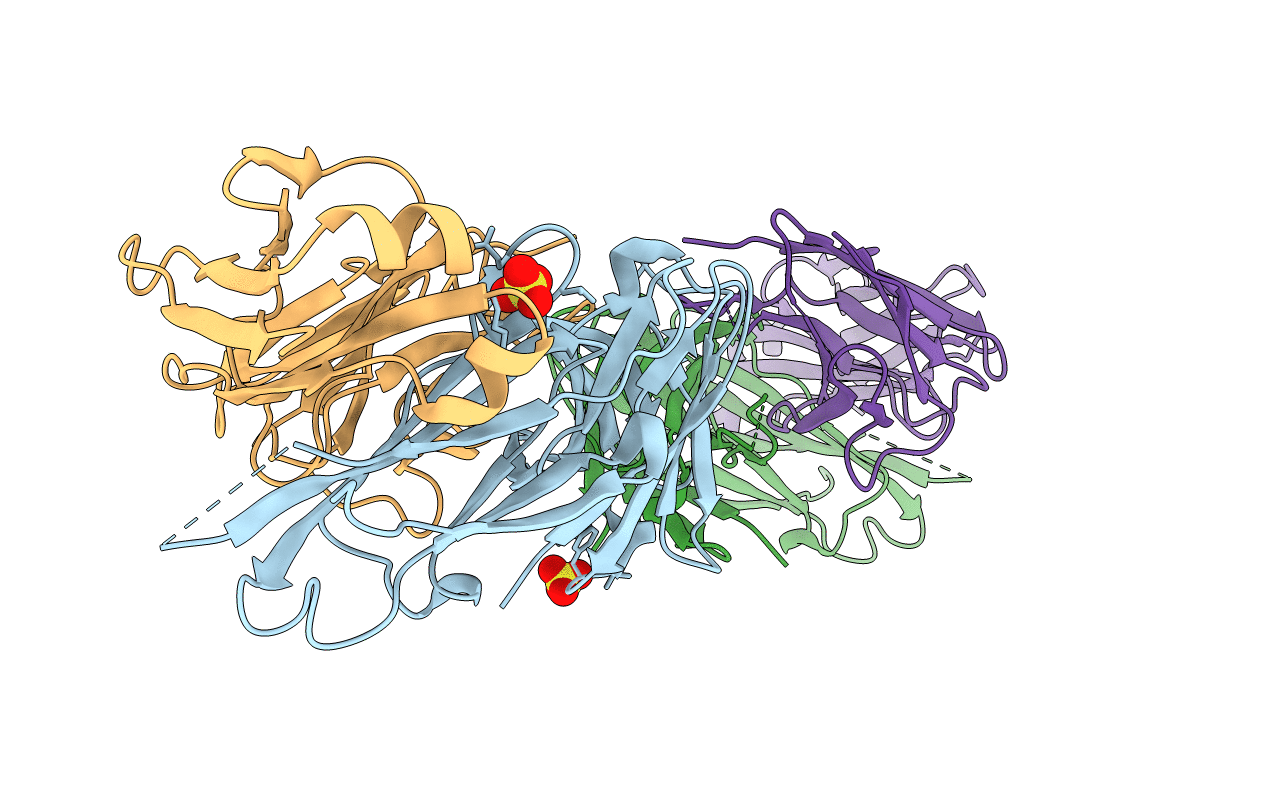
PDB ID:
2ORB
Keywords:
Title:
The structure of the anti-c-myc antibody 9E10 Fab fragment
Biological Source:
Source Organism(s):
Mus musculus (Taxon ID: 10090)
Method Details:
Experimental Method:
Resolution:
2.20 Å
R-Value Free:
0.31
R-Value Work:
0.26
Space Group:
P 1 21 1
