Deposition Date
2007-02-01
Release Date
2007-02-20
Last Version Date
2024-10-30
Entry Detail
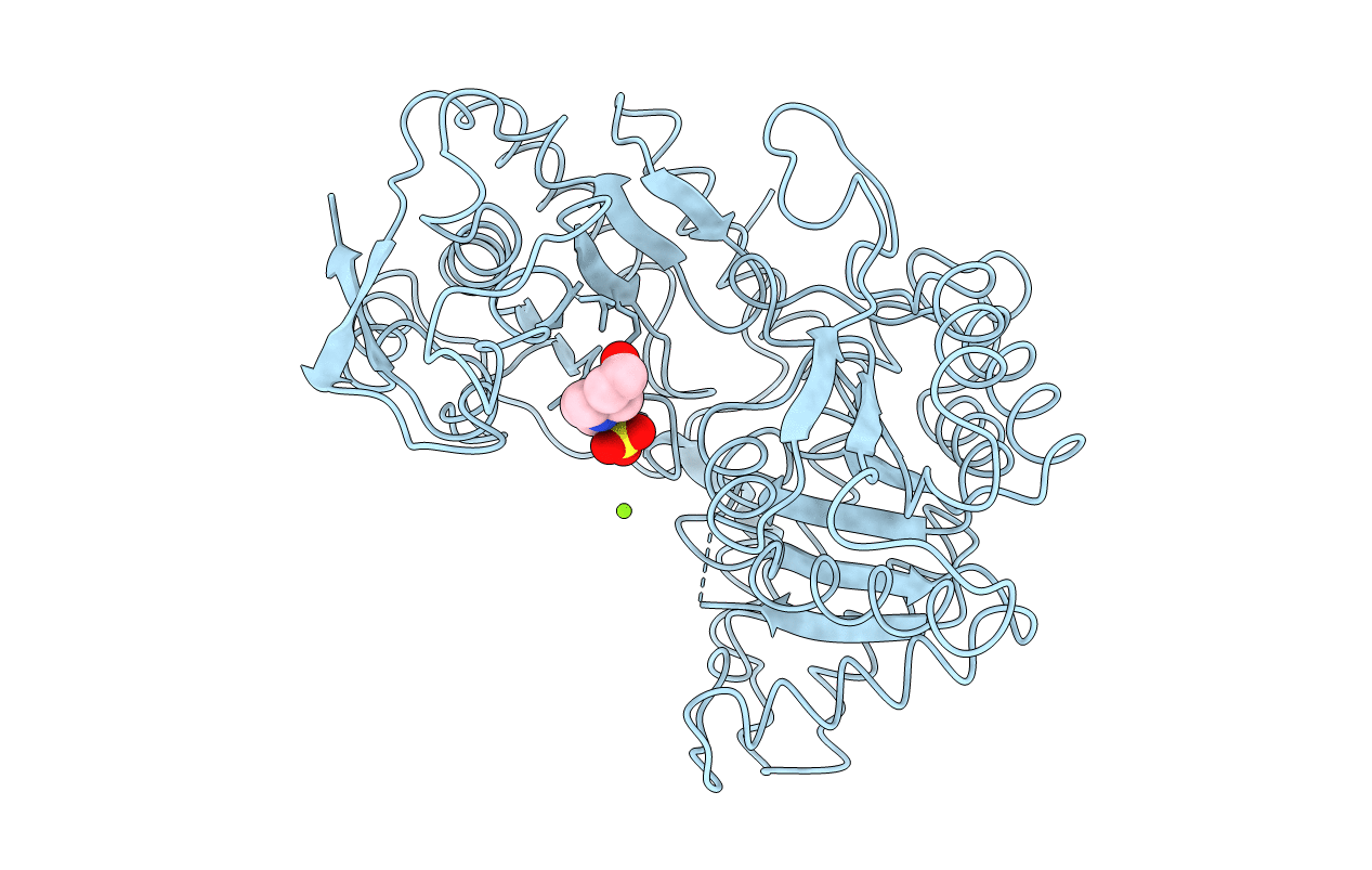
PDB ID:
2OQX
Keywords:
Title:
Crystal Structure of the apo form of E. coli tryptophanase at 1.9 A resolution
Biological Source:
Source Organism(s):
Escherichia coli (Taxon ID: 562)
Method Details:
Experimental Method:
Resolution:
1.90 Å
R-Value Free:
0.23
R-Value Work:
0.20
R-Value Observed:
0.21
Space Group:
F 2 2 2