2OQS image
Deposition Date 2007-02-01
Release Date 2007-09-04
Last Version Date 2023-12-27
Entry Detail
PDB ID:
2OQS
Title:
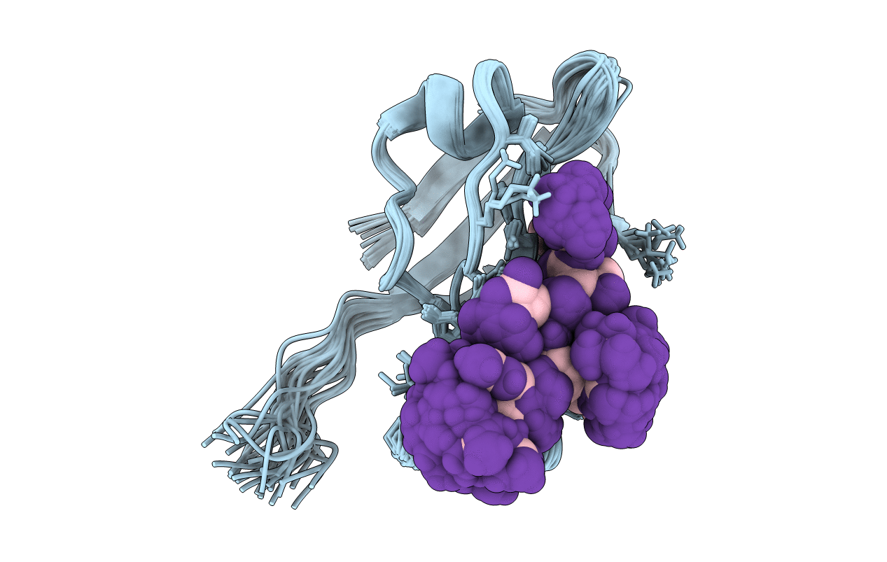
Structure of the hDLG/SAP97 PDZ2 in complex with HPV-18 papillomavirus E6 peptide
Biological Source:
Source Organism:
Homo sapiens (Taxon ID: 9606)
(Taxon ID: )
Method Details:
Experimental Method:
Conformers Calculated:
30
Conformers Submitted:
30
Selection Criteria:
all calculated structures submitted
Macromolecular Entities
Polymer Type:polypeptide(L)
Molecule:Disks large homolog 1
Gene (Uniprot):DLG1
Chain IDs:A
Chain Length:97
Number of Molecules:1
Biological Source:Homo sapiens
Polymer Type:polypeptide(L)
Molecule:C-terminal HPV-18 E6 peptide
Chain IDs:B
Chain Length:6
Number of Molecules:1
Biological Source:
Ligand Molecules
Primary Citation
Solution structure of the hDlg/SAP97 PDZ2 domain and its mechanism of interaction with HPV-18 papillomavirus E6 protein.
Biochemistry 46 10864 10874 (2007)
PMID: 17713926 DOI: 10.1021/bi700879k

Abstact

The E6 protein from high-risk types of human papillomavirus (HPV) binds PDZ-domain containing proteins and targets them for degradation. We used isothermal titration calorimetry to measure the interaction of a peptide from the C-terminus of HPV-18 E6 to the second PDZ domain (PDZ2) from the human homologue of the Drosophila discs large tumor suppressor protein (hDlg). Isothermal titration calorimetry experiments with a series of peptides showed that HPV-18 E6 bound hDlg PDZ2 about 5-fold stronger than HPV-16 E6, that the contribution of Arg154 to binding was about 1 kcal/mol, and that the binding was disabled by phosphorylation at Thr156. We then used NMR to determine the solution structure of the complex of PDZ2 bound to the HPV-18 E6 peptide. The resultant structures were of high quality and had backbone root-mean-square deviations of less than 0.5 A. The structure shows a novel mode of interaction in which six residues of the HPV-18 E6 peptide are contacted by the PDZ2 domain, in contrast to the typical four residues used by class I PDZ domains. Molecular dynamics simulations supported a model in which the C- and N-terminal ends of the peptide had different mobilities within the complex. Comparison of the NMR complex structure to previously determined X-ray structures of PDZ2 by itself and bound to different peptides allows a description of conformational changes required for PDZ2 to bind to HPV-18 E6.

Legend

Protein

Chemical

Disease

Primary Citation of related structures