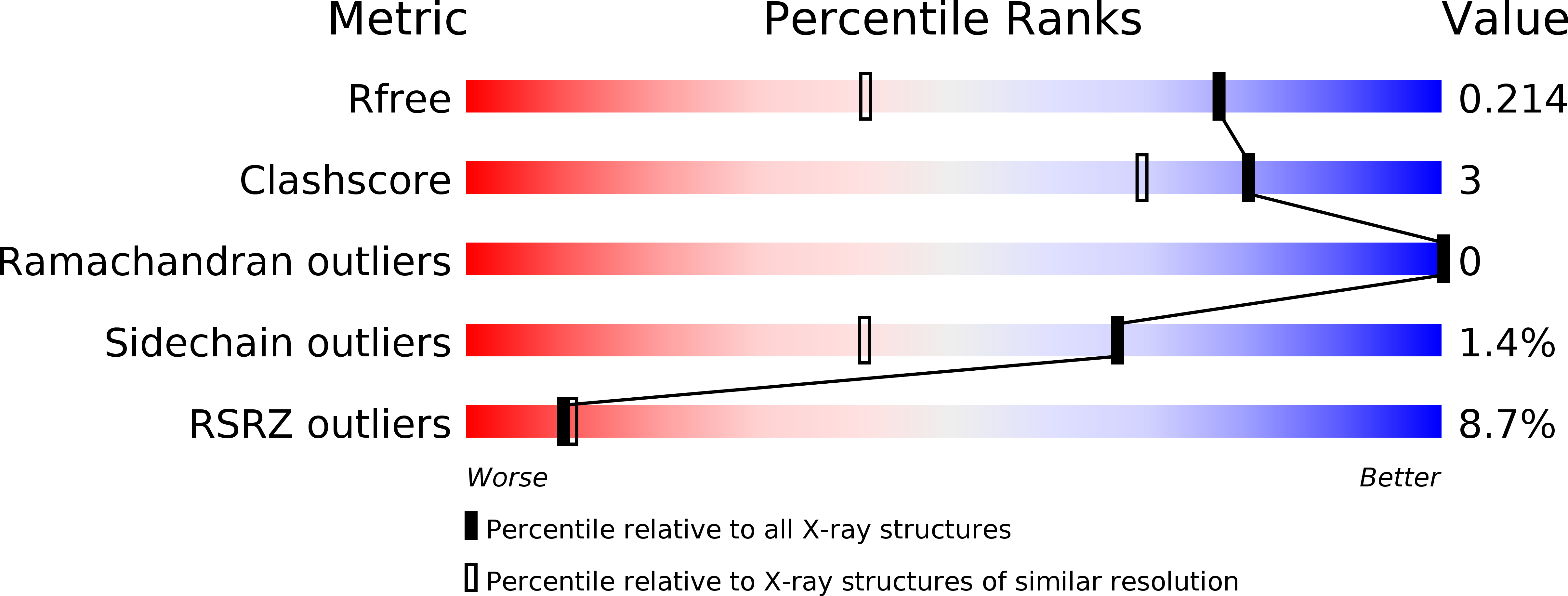
2OOC image
Deposition Date 2007-01-25
Release Date 2007-02-06
Last Version Date 2024-10-30
Entry Detail
PDB ID:
2OOC
Keywords:
Title:
Crystal structure of Histidine Phosphotransferase ShpA (NP_419930.1) from Caulobacter crescentus at 1.52 A resolution
Biological Source:
Source Organism:
Host Organism:
Method Details:
Experimental Method:
Resolution:
1.52 Å
R-Value Free:
0.20
R-Value Work:
0.16
R-Value Observed:
0.16
Space Group:
P 32 2 1
Macromolecular Entities
Structures with similar UniProt ID
Protein Blast
Polymer Type:polypeptide(L)
Molecule:Histidine phosphotransferase
Chain IDs:A, B
Chain Length:113
Number of Molecules:2
Biological Source:Caulobacter crescentus
Modified Residue
Compound ID Chain ID Parent Comp ID Details 2D Image
MSE A MET SELENOMETHIONINE
Primary Citation

Abstact

Cell-cycle-regulated stalk biogenesis in Caulobacter crescentus is controlled by a multistep phosphorelay system consisting of the hybrid histidine kinase ShkA, the histidine phosphotransfer (HPt) protein ShpA, and the response regulator TacA. ShpA shuttles phosphoryl groups between ShkA and TacA. When phosphorylated, TacA triggers a downstream transcription cascade for stalk synthesis in an RpoN-dependent manner. The crystal structure of ShpA was determined to 1.52 A resolution. ShpA belongs to a family of monomeric HPt proteins that feature a highly conserved four-helix bundle. The phosphorylatable histidine His56 is located on the surface of the helix bundle and is fully solvent exposed. One end of the four-helix bundle in ShpA is shorter compared with other characterized HPt proteins, whereas the face that potentially interacts with the response regulators is structurally conserved. Similarities of the interaction surface around the phosphorylation site suggest that ShpA is likely to share a common mechanism for molecular recognition and phosphotransfer with yeast phosphotransfer protein YPD1 despite their low overall sequence similarity.

Legend

Protein

Chemical

Disease

Primary Citation of related structures