Planned Maintenance: Some services may turn out to be unavailable from 15th January, 2026 to 16th January, 2026. We apologize for the inconvenience!

2OLG image
Deposition Date 2007-01-19
Release Date 2007-02-06
Last Version Date 2024-11-20
Entry Detail
PDB ID:
2OLG
Keywords:
Title:
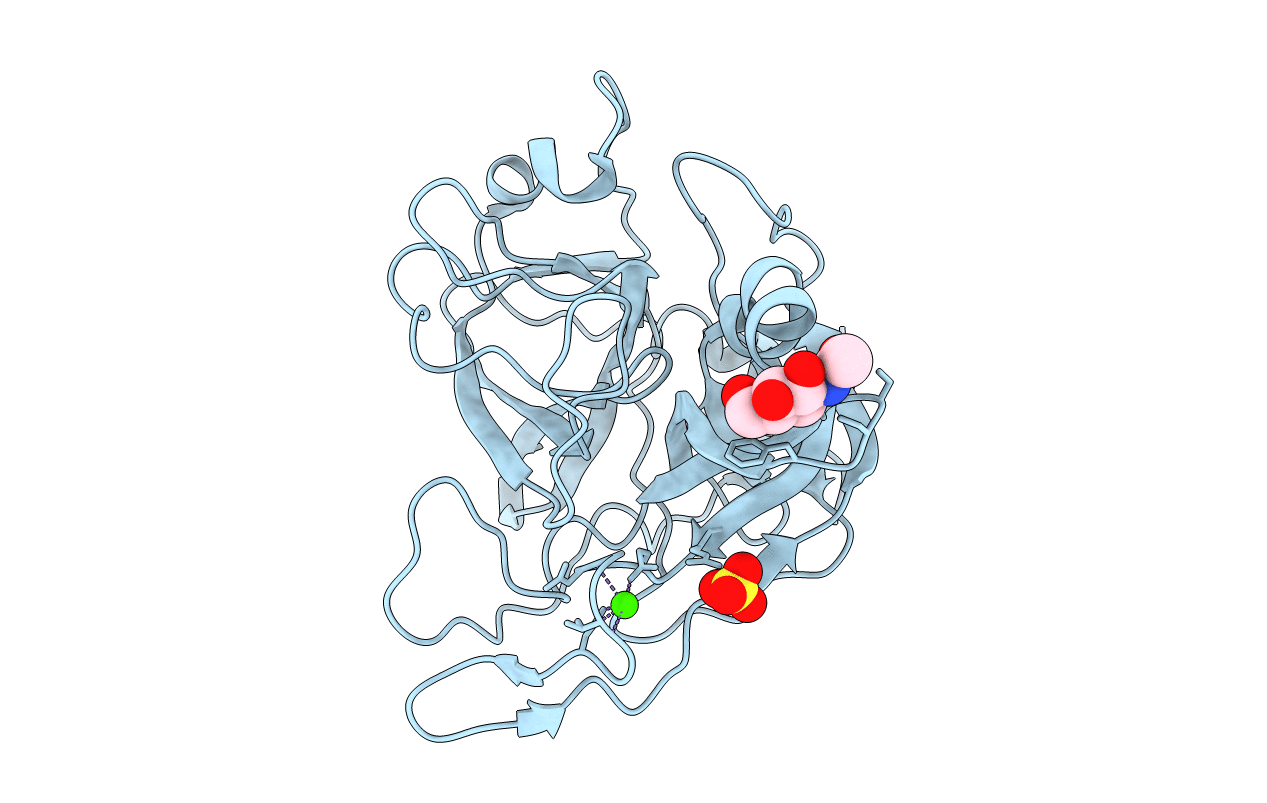
Crystal structure of the serine protease domain of prophenoloxidase activating factor-I in a zymogen form
Biological Source:
Source Organism(s):
Expression System(s):
Method Details:
Experimental Method:
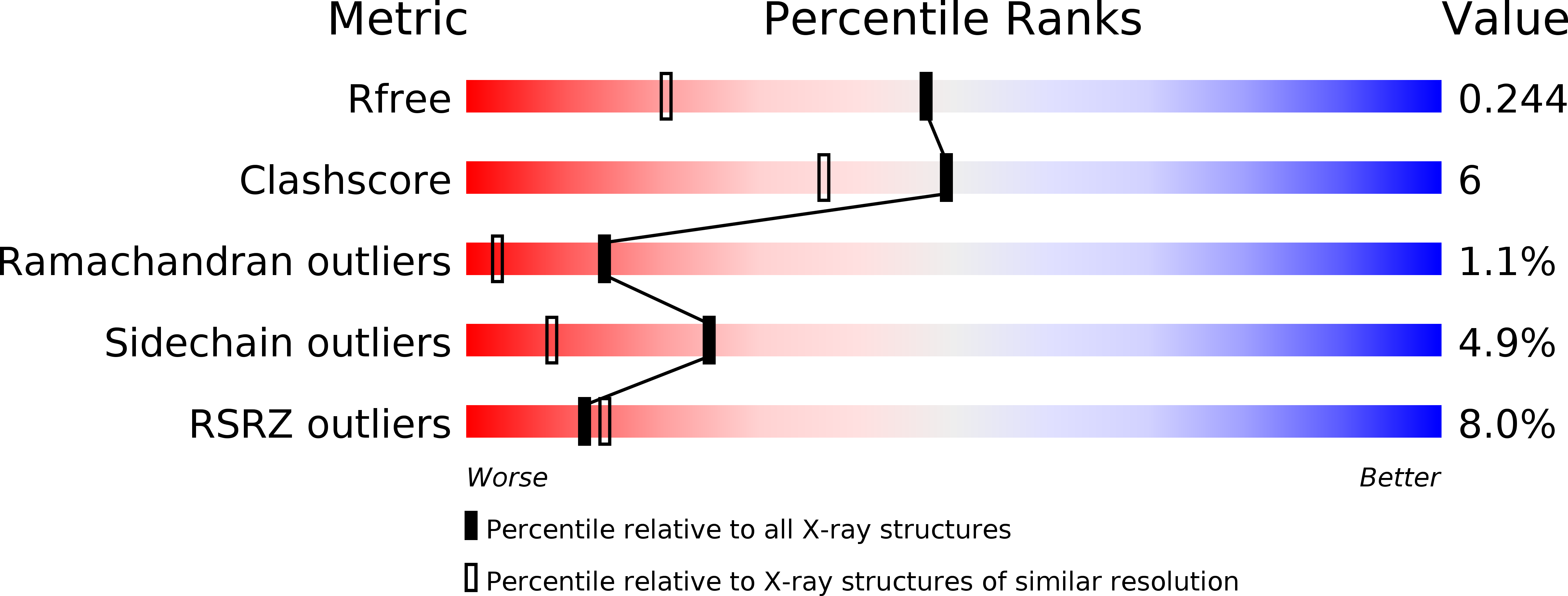
Resolution:
1.70 Å
R-Value Free:
0.24
R-Value Work:
0.20
R-Value Observed:
0.20
Space Group:
P 21 21 21
Macromolecular Entities
Structures with similar UniProt ID
Protein Blast
Polymer Type:polypeptide(L)
Molecule:Pro-phenoloxidase activating enzyme-I
Gene (Uniprot):PPAF1
Chain IDs:A
Chain Length:278
Number of Molecules:1
Biological Source:Holotrichia diomphalia
Modified Residue
Compound ID Chain ID Parent Comp ID Details 2D Image
ASN A ASN GLYCOSYLATION SITE
Primary Citation
Crystal structure of the serine protease domain of prophenoloxidase activating factor-I
J.Biol.Chem. 282 10783 10791 (2007)
PMID: 17287215 DOI: 10.1074/jbc.M611556200

Abstact

A family of serine proteases (SPs) mediates the proteolytic cascades of embryonic development and immune response in invertebrates. These proteases, called easter-type SPs, consist of clip and chymotrypsin-like SP domains. The SP domain of easter-type proteases differs from those of typical SPs in its primary structure. Herein, we report the first crystal structure of the SP domain of easter-type proteases, presented as that of prophenoloxidase activating factor (PPAF)-I in zymogen form. This structure reveals several important structural features including a bound calcium ion, an additional loop with a unique disulfide linkage, a canyon-like deep active site, and an exposed activation loop. We subsequently show the role of the bound calcium and the proteolytic susceptibility of the activation loop, which occurs in a clip domain-independent manner. Based on biochemical study in the presence of heparin, we suggest that PPAF-III, highly homologous to PPAF-I, contains a surface patch that is responsible for enhancing the catalytic activity through interaction with a nonsubstrate region of a target protein. These results provide insights into an activation mechanism of easter-type proteases in proteolytic cascades, in comparison with the well studied blood coagulation enzymes in mammals.

Legend

Protein

Chemical

Disease

Primary Citation of related structures
Feedback Form
Name
Email
Institute
Feedback