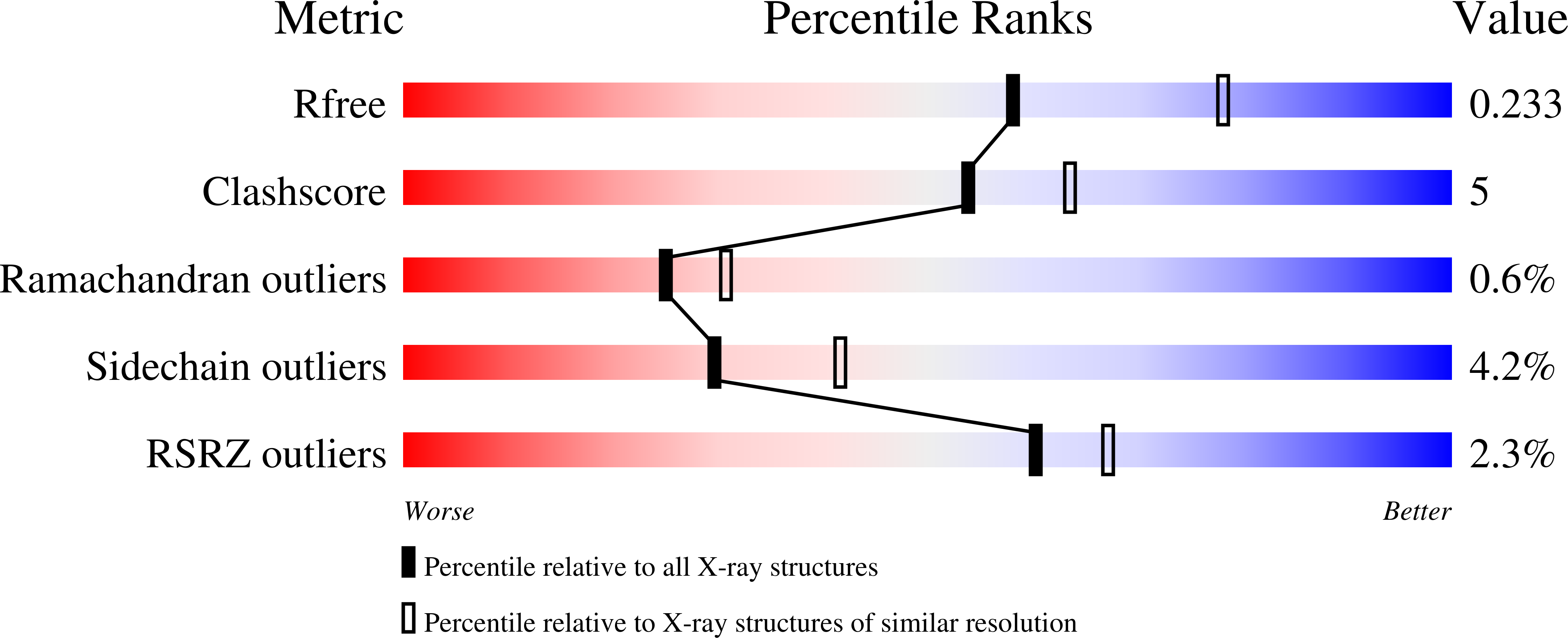
2OK5 image
Deposition Date 2007-01-16
Release Date 2007-02-27
Last Version Date 2024-11-20
Entry Detail
PDB ID:
2OK5
Keywords:
Title:
Human Complement factor B
Biological Source:
Source Organism:
Homo sapiens (Taxon ID: 9606)
Host Organism:
Method Details:
Experimental Method:
Resolution:
2.30 Å
R-Value Free:
0.24
R-Value Work:
0.19
Space Group:
P 31 2 1
Macromolecular Entities
Structures with similar UniProt ID
Protein Blast
Polymer Type:polypeptide(L)
Molecule:Complement factor B
Gene (Uniprot):CFB
Chain IDs:A
Chain Length:752
Number of Molecules:1
Biological Source:Homo sapiens
Modified Residue
Compound ID Chain ID Parent Comp ID Details 2D Image
ASN A ASN GLYCOSYLATION SITE
Primary Citation
Factor B structure provides insights into activation of the central protease of the complement system.
Nat.Struct.Mol.Biol. 14 224 228 (2007)
PMID: 17310251 DOI: 10.1038/nsmb1210

Abstact

Factor B is the central protease of the complement system of immune defense. Here, we present the crystal structure of human factor B at 2.3-A resolution, which reveals how the five-domain proenzyme is kept securely inactive. The canonical activation helix of the Von Willebrand factor A (VWA) domain is displaced by a helix from the preceding domain linker. The two helices conformationally link the scissile-activation peptide and the metal ion-dependent adhesion site required for binding of the ligand C3b. The data suggest that C3b binding displaces the three N-terminal control domains and reshuffles the two central helices. Reshuffling of the helices releases the scissile bond for final proteolytic activation and generates a new interface between the VWA domain and the serine protease domain. This allosteric mechanism is crucial for tight regulation of the complement-amplification step in the immune response.

Legend

Protein

Chemical

Disease

Primary Citation of related structures