Deposition Date
2007-01-12
Release Date
2007-09-25
Last Version Date
2023-11-15
Entry Detail
PDB ID:
2OJK
Keywords:
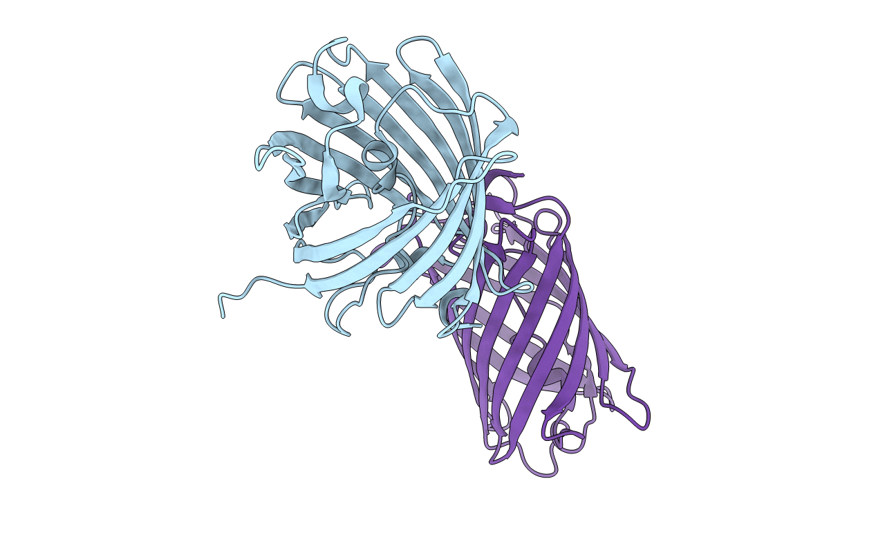
Title:
Crystal Structure of Green Fluorescent Protein from Zoanthus sp at 2.2 A Resolution
Biological Source:
Source Organism(s):
Zoanthus sp. (Taxon ID: 105402)
Expression System(s):
Method Details:
Experimental Method:
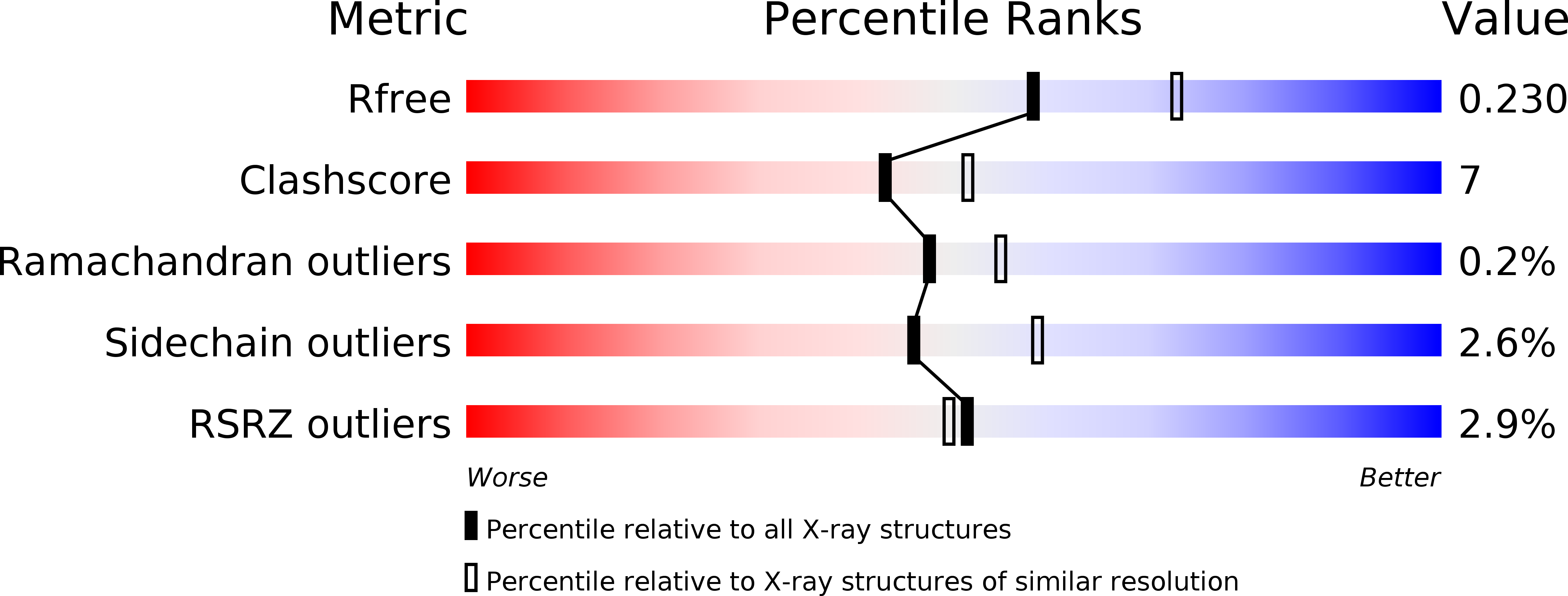
Resolution:
2.20 Å
R-Value Free:
0.23
R-Value Work:
0.18
R-Value Observed:
0.18
Space Group:
P 62 2 2