2OGG image
Deposition Date 2007-01-05
Release Date 2007-02-06
Last Version Date 2024-10-09
Entry Detail
PDB ID:
2OGG
Keywords:
Title:
Structure of B. subtilis trehalose repressor (TreR) effector binding domain
Biological Source:
Source Organism:
Method Details:
Experimental Method:
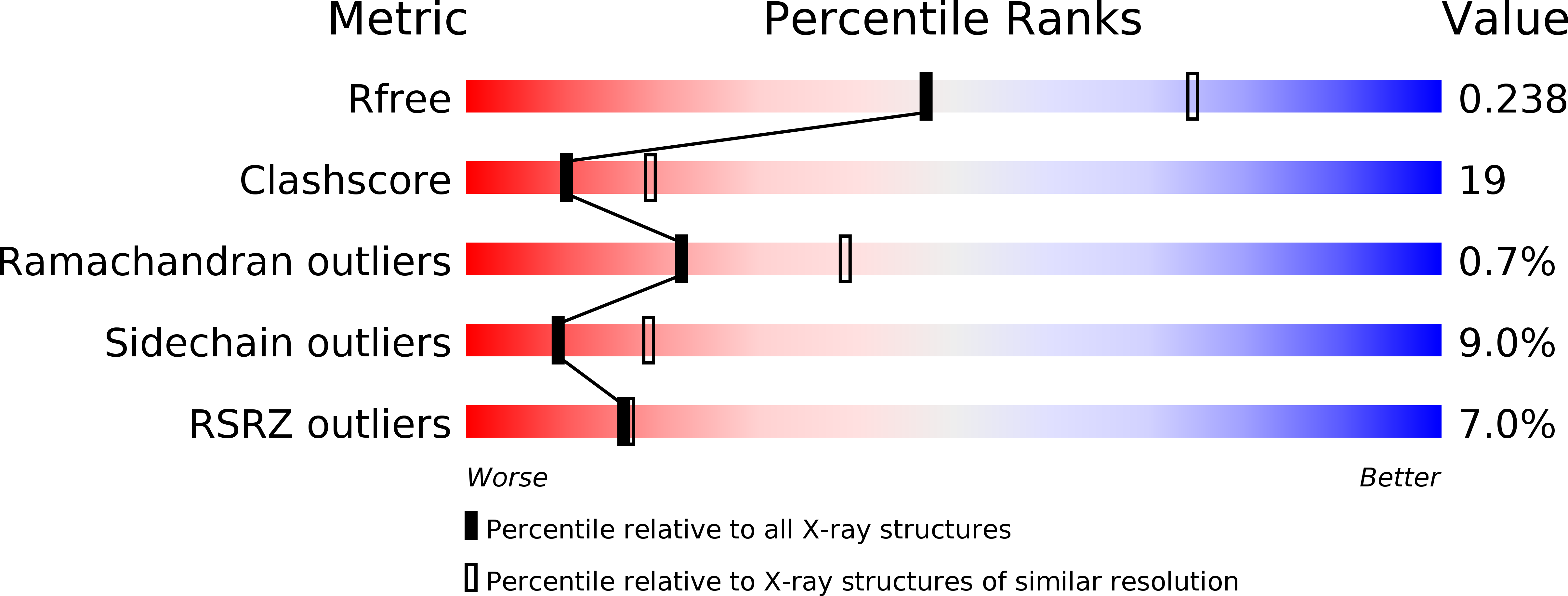
Resolution:
2.50 Å
R-Value Free:
0.24
R-Value Work:
0.19
R-Value Observed:
0.19
Space Group:
I 41 2 2
Macromolecular Entities
Structures with similar UniProt ID
Protein Blast
Polymer Type:polypeptide(L)
Molecule:Trehalose operon transcriptional repressor
Gene (Uniprot):treR
Chain IDs:A
Chain Length:152
Number of Molecules:1
Biological Source:Bacillus subtilis
Modified Residue
Compound ID Chain ID Parent Comp ID Details 2D Image
MSE A MET SELENOMETHIONINE
Primary Citation
The crystal structure of the effector-binding domain of the trehalose repressor TreR from Bacillus subtilis 168 reveals a unique quarternary assembly.
Proteins 69 679 682 (2007)
PMID: 17705272 DOI: 10.1002/prot.21516
Primary Citation of related structures