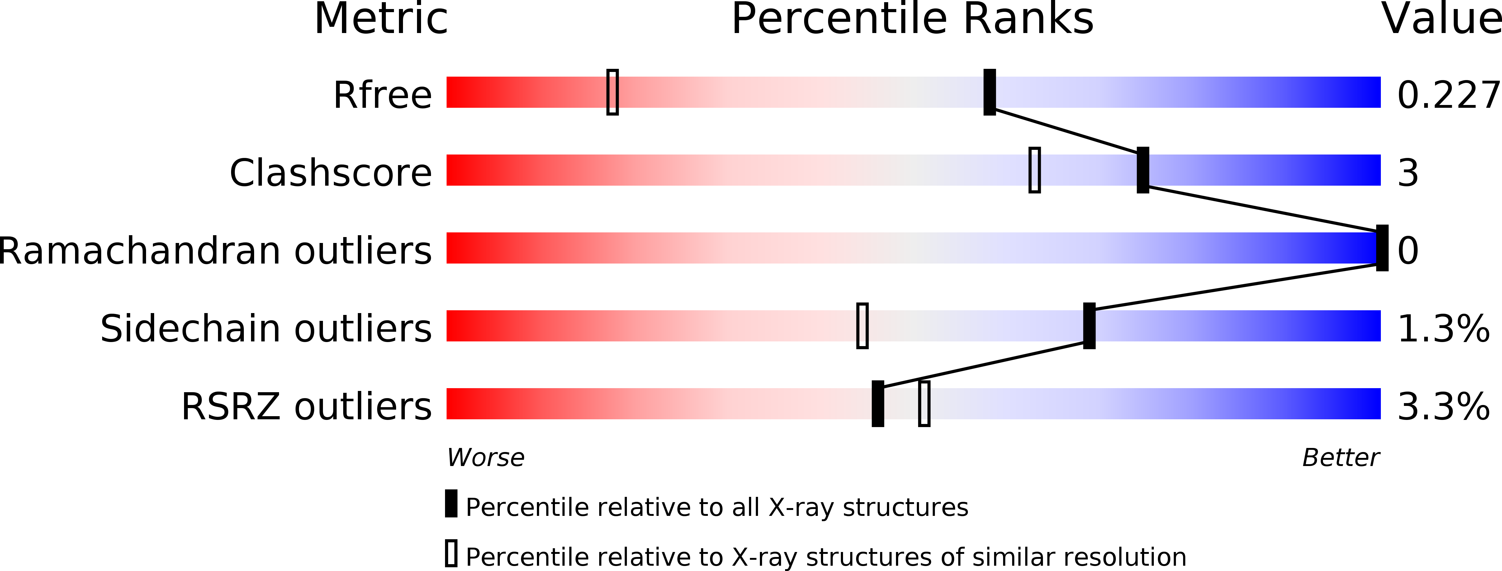
2OFK image
Deposition Date 2007-01-03
Release Date 2007-05-15
Last Version Date 2023-12-27
Entry Detail
PDB ID:
2OFK
Keywords:
Title:
Crystal Structure of 3-methyladenine DNA glycosylase I (TAG)
Biological Source:
Source Organism:
Salmonella typhi (Taxon ID: 601)
Host Organism:
Method Details:
Experimental Method:
Resolution:
1.50 Å
R-Value Free:
0.19
R-Value Work:
0.16
R-Value Observed:
0.16
Space Group:
P 1 21 1
Macromolecular Entities
Polymer Type:polypeptide(L)
Molecule:3-methyladenine DNA glycosylase I, constitutive
Gene (Uniprot):tag
Chain IDs:A, B
Chain Length:183
Number of Molecules:2
Biological Source:Salmonella typhi
Primary Citation
DNA damage recognition and repair by 3-methyladenine DNA glycosylase I (TAG).
Embo J. 26 2411 2420 (2007)
PMID: 17410210 DOI: 10.1038/sj.emboj.7601649

Abstact

DNA glycosylases help maintain the genome by excising chemically modified bases from DNA. Escherichia coli 3-methyladenine DNA glycosylase I (TAG) specifically catalyzes the removal of the cytotoxic lesion 3-methyladenine (3mA). The molecular basis for the enzymatic recognition and removal of 3mA from DNA is currently a matter of speculation, in part owing to the lack of a structure of a 3mA-specific glycosylase bound to damaged DNA. Here, high-resolution crystal structures of Salmonella typhi TAG in the unliganded form and in a ternary product complex with abasic DNA and 3mA nucleobase are presented. Despite its structural similarity to the helix-hairpin-helix superfamily of DNA glycosylases, TAG has evolved a modified strategy for engaging damaged DNA. In contrast to other glycosylase-DNA structures, the abasic ribose is not flipped into the TAG active site. This is the first structural demonstration that conformational relaxation must occur in the DNA upon base hydrolysis. Together with mutational studies of TAG enzymatic activity, these data provide a model for the specific recognition and hydrolysis of 3mA from DNA.

Legend

Protein

Chemical

Disease

Primary Citation of related structures