Deposition Date
2006-12-27
Release Date
2008-01-22
Last Version Date
2024-10-30
Entry Detail
PDB ID:
2ODY
Keywords:
Title:
Thrombin-bound boophilin displays a functional and accessible reactive-site loop
Biological Source:
Source Organism(s):
Rhipicephalus microplus (Taxon ID: 6941)
Bos taurus (Taxon ID: 9913)
Bos taurus (Taxon ID: 9913)
Expression System(s):
Method Details:
Experimental Method:
Resolution:
2.35 Å
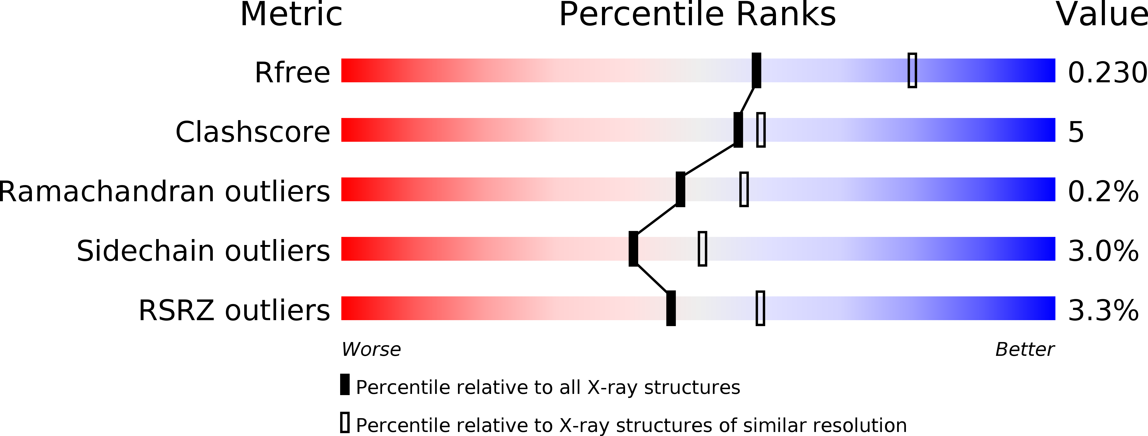
R-Value Free:
0.22
R-Value Work:
0.19
R-Value Observed:
0.19
Space Group:
P 21 21 21