2ODJ image
Deposition Date 2006-12-22
Release Date 2007-10-16
Last Version Date 2024-10-30
Entry Detail
PDB ID:
2ODJ
Title:
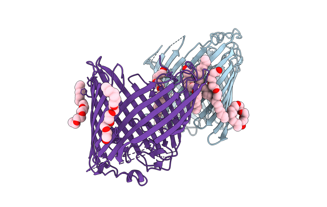
Crystal structure of the outer membrane protein OprD from Pseudomonas aeruginosa
Biological Source:
Source Organism:
Host Organism:
Method Details:
Experimental Method:
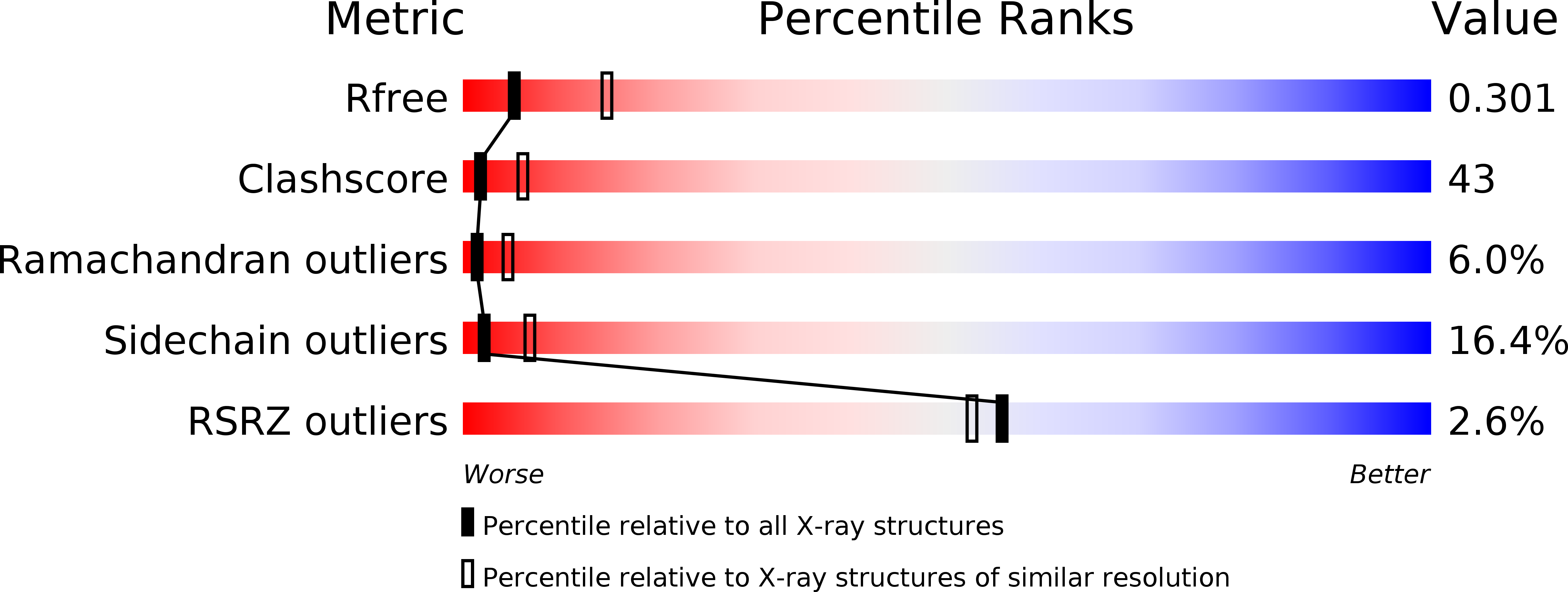
Resolution:
2.90 Å
R-Value Free:
0.31
R-Value Work:
0.24
R-Value Observed:
0.24
Space Group:
C 1 2 1
Macromolecular Entities
Structures with similar UniProt ID
Protein Blast
Polymer Type:polypeptide(L)
Molecule:Porin D
Gene (Uniprot):oprD
Chain IDs:A, B
Chain Length:428
Number of Molecules:2
Biological Source:Pseudomonas aeruginosa
Modified Residue
Compound ID Chain ID Parent Comp ID Details 2D Image
MSE A MET SELENOMETHIONINE
Ligand Molecules
Primary Citation
Structural insight into OprD substrate specificity.
Nat.Struct.Mol.Biol. 14 1108 1109 (2007)
PMID: 17952093 DOI: 10.1038/nsmb1304

Abstact

OprD proteins form a large family of substrate-specific outer-membrane channels in Gram-negative bacteria. We report here the X-ray crystal structure of OprD from Pseudomonas aeruginosa, which reveals a monomeric 18-stranded beta-barrel characterized by a very narrow pore constriction, with a positively charged basic ladder on one side and an electronegative pocket on the other side. The location of highly conserved residues in OprD suggests that the structure represents the general architecture of OprD channels.

Legend

Protein

Chemical

Disease

Primary Citation of related structures