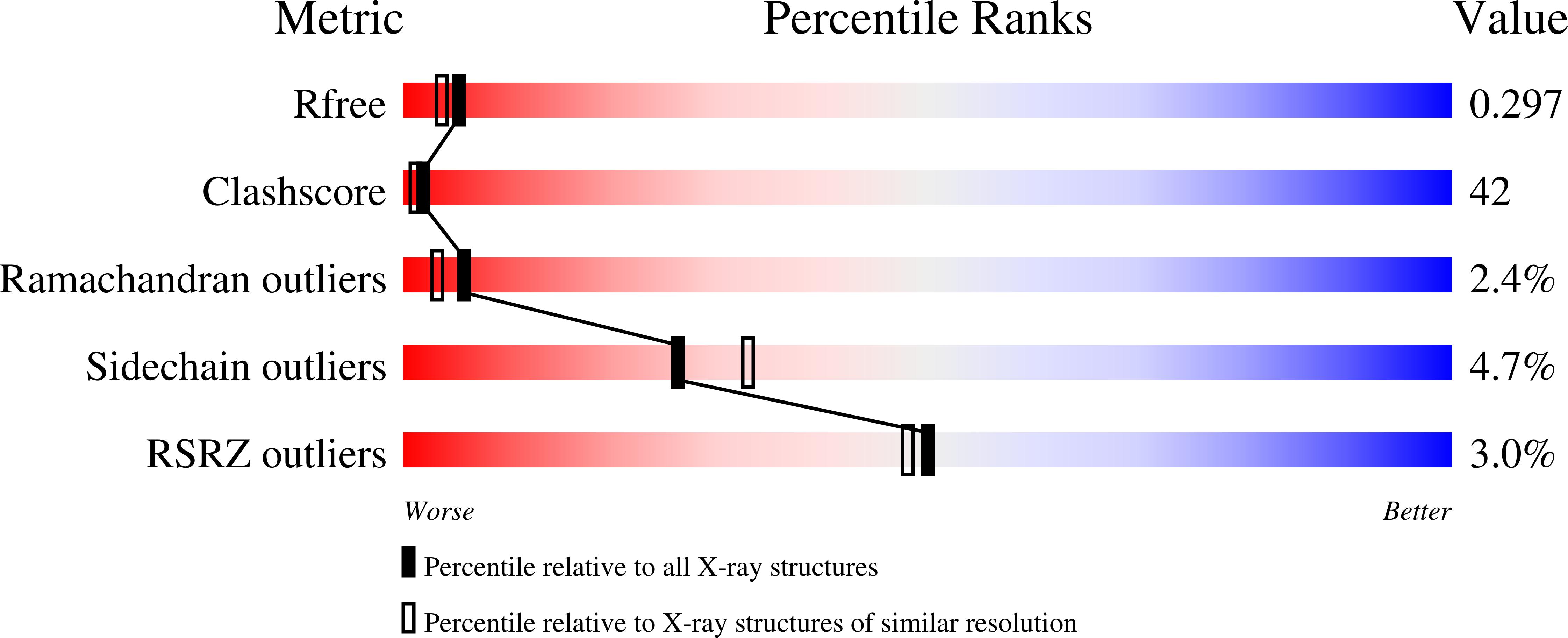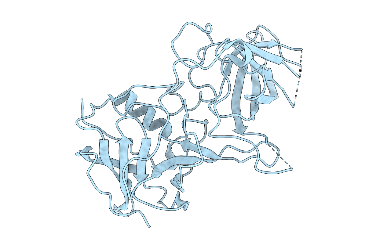
2OBR image
Deposition Date 2006-12-20
Release Date 2007-04-24
Last Version Date 2023-10-25
Entry Detail
PDB ID:
2OBR
Keywords:
Title:
Crystal Structures of P Domain of Norovirus VA387
Biological Source:
Source Organism:
Norovirus (Taxon ID: 142786)
Method Details:
Experimental Method:
Resolution:
2.20 Å
R-Value Free:
0.29
R-Value Work:
0.22
R-Value Observed:
0.23
Space Group:
C 2 2 21
Macromolecular Entities
Structures with similar UniProt ID
Protein Blast
Polymer Type:polypeptide(L)
Molecule:Capsid protein
Mutagens:T355S,F375L
Chain IDs:A
Chain Length:327
Number of Molecules:1
Biological Source:Norovirus
Primary Citation
Structural basis for the recognition of blood group trisaccharides by norovirus.
J.Virol. 81 5949 5957 (2007)
PMID: 17392366 DOI: 10.1128/JVI.00219-07

Abstact

Noroviruses are one of the major causes of nonbacterial gastroenteritis epidemics in humans. Recent studies on norovirus receptors show that different noroviruses recognize different human histo-blood group antigens (HBGAs), and eight receptor binding patterns of noroviruses have been identified. The P domain of the norovirus capsids is directly involved in this recognition. To determine the precise locations and receptor binding modes of HBGA carbohydrates on the viral capsids, a recombinant P protein of a GII-4 strain norovirus, VA387, was cocrystallized with synthetic type A or B trisaccharides. Based on complex crystal structures observed at a 2.0-A resolution, we demonstrated that the receptor binding site lies at the outermost end of the P domain and forms an extensive hydrogen-bonding network with the saccharide ligand. The A and B trisaccharides display similar binding modes, and the common fucose ring plays a key role in this interaction. The extensive interface between the two protomers in a P dimer also plays a crucial role in the formation of the receptor binding interface.

Legend

Protein

Chemical

Disease

Primary Citation of related structures