2OA0 image
Deposition Date 2006-12-14
Release Date 2007-02-06
Last Version Date 2023-08-30
Entry Detail
PDB ID:
2OA0
Keywords:
Title:
Crystal structure of Calcium ATPase with bound ADP and cyclopiazonic acid
Biological Source:
Source Organism:
Method Details:
Experimental Method:
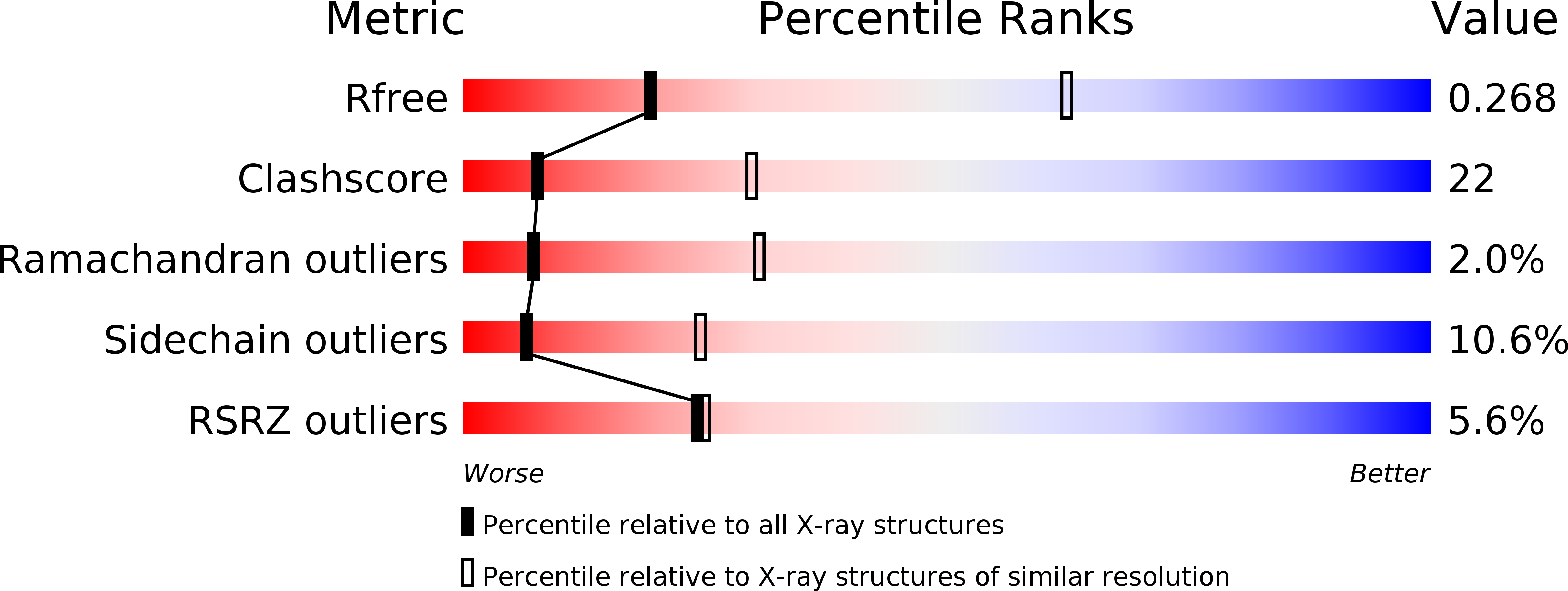
Resolution:
3.40 Å
R-Value Free:
0.32
R-Value Work:
0.29
R-Value Observed:
0.29
Space Group:
P 1 21 1
Macromolecular Entities
Polymer Type:polypeptide(L)
Molecule:Sarcoplasmic/endoplasmic reticulum calcium ATPase 1
Gene (Uniprot):ATP2A1
Chain IDs:A
Chain Length:994
Number of Molecules:1
Biological Source:Oryctolagus cuniculus
Primary Citation
The molecular basis for cyclopiazonic Acid inhibition of the sarcoplasmic reticulum calcium pump.
J.Biol.Chem. 282 9748 9757 (2007)
PMID: 17259168 DOI: 10.1074/jbc.M611653200

Abstact

The sarcoplasmic reticulum Ca(2+)-ATPase is essential for calcium reuptake in the muscle contraction-relaxation cycle. Here we present structures of a calcium-free state with bound cyclopiazonic acid (CPA) and magnesium fluoride at 2.65 A resolution and a calcium-free state with bound CPA and ADP at 3.4A resolution. In both structures, CPA occupies the calcium access channel delimited by transmembrane segments M1-M4. Inhibition of Ca(2+)-ATPase is stabilized by a polar pocket that surrounds the tetramic acid of CPA and a hydrophobic platform that cradles the inhibitor. The calcium pump residues involved include Gln(56), Leu(61), Val(62), and Asn(101). We conclude that CPA inhibits the calcium pump by blocking the calcium access channel and immobilizing a subset of transmembrane helices. In the E2(CPA) structure, ADP is bound in a distinct orientation within the nucleotide binding pocket. The adenine ring is sandwiched between Arg(489) of the nucleotide-binding domain and Arg(678) of the phosphorylation domain. This mode of binding conforms to an adenine recognition motif commonly found in ATP-dependent proteins.

Legend

Protein

Chemical

Disease

Primary Citation of related structures