Deposition Date
2006-12-12
Release Date
2007-03-27
Last Version Date
2024-10-30
Entry Detail
PDB ID:
2O8V
Keywords:
Title:
PAPS reductase in a covalent complex with thioredoxin C35A
Biological Source:
Source Organism(s):
Escherichia coli (Taxon ID: 562)
Expression System(s):
Method Details:
Experimental Method:
Resolution:
3.00 Å
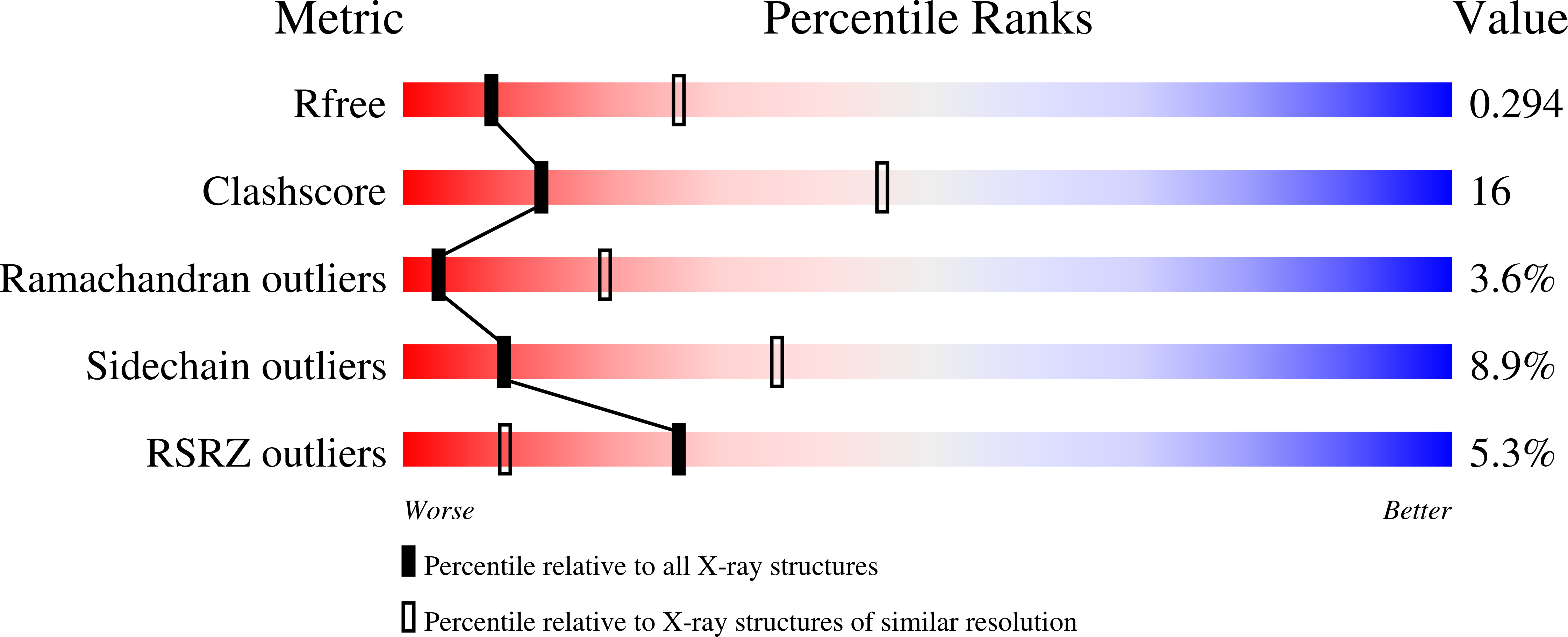
R-Value Free:
0.30
R-Value Work:
0.29
R-Value Observed:
0.29
Space Group:
C 2 2 21