Deposition Date
2006-12-12
Release Date
2007-05-01
Last Version Date
2024-10-30
Entry Detail
PDB ID:
2O88
Keywords:
Title:
Crystal structure of the N114A mutant of ABL-SH3 domain complexed with a designed high-affinity peptide ligand: implications for SH3-ligand interactions
Biological Source:
Source Organism(s):
Homo sapiens (Taxon ID: 9606)
synthetic construct (Taxon ID: 32630)
synthetic construct (Taxon ID: 32630)
Expression System(s):
Method Details:
Experimental Method:
Resolution:
1.75 Å
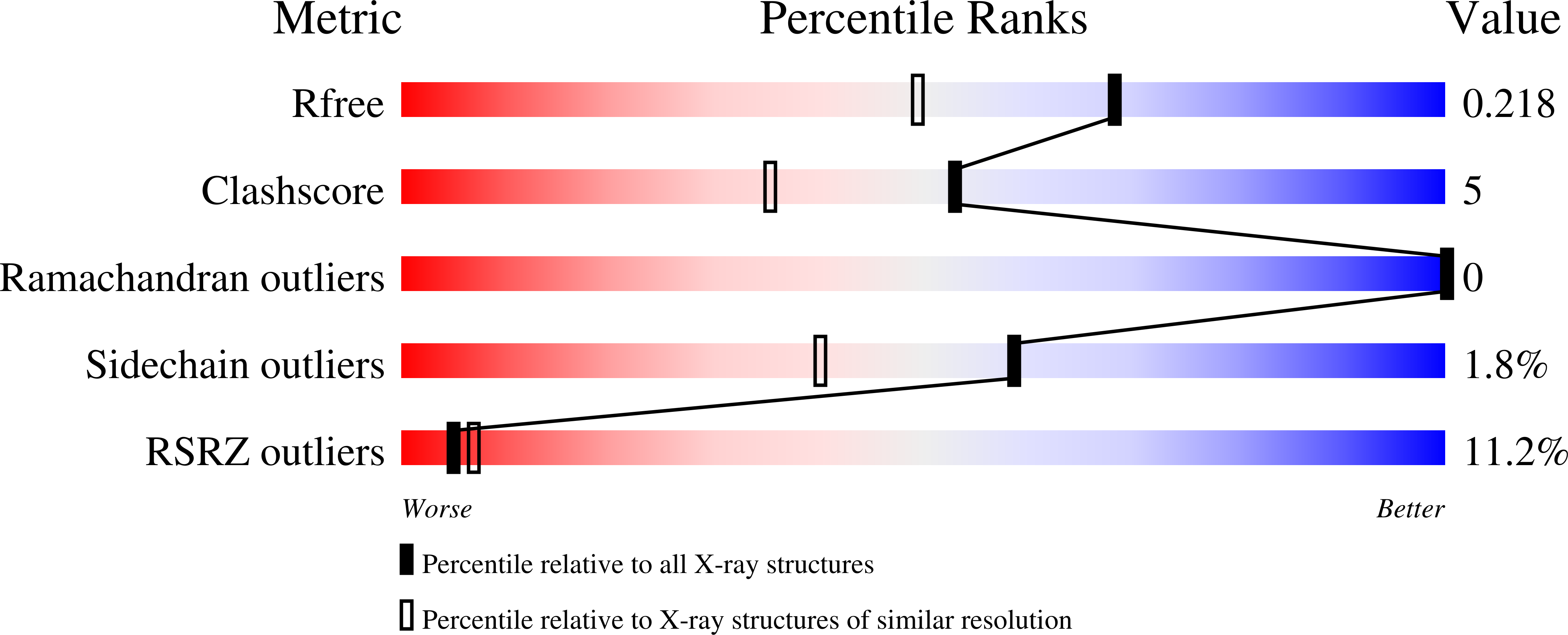
R-Value Free:
0.21
R-Value Work:
0.16
R-Value Observed:
0.17
Space Group:
P 21 21 21