2O7O image
Deposition Date 2006-12-11
Release Date 2007-05-15
Last Version Date 2023-10-25
Entry Detail
PDB ID:
2O7O
Title:
Crystal structure analysis of TetR(D) complex with doxycycline
Biological Source:
Source Organism:
Escherichia coli (Taxon ID: 562)
Host Organism:
Method Details:
Experimental Method:
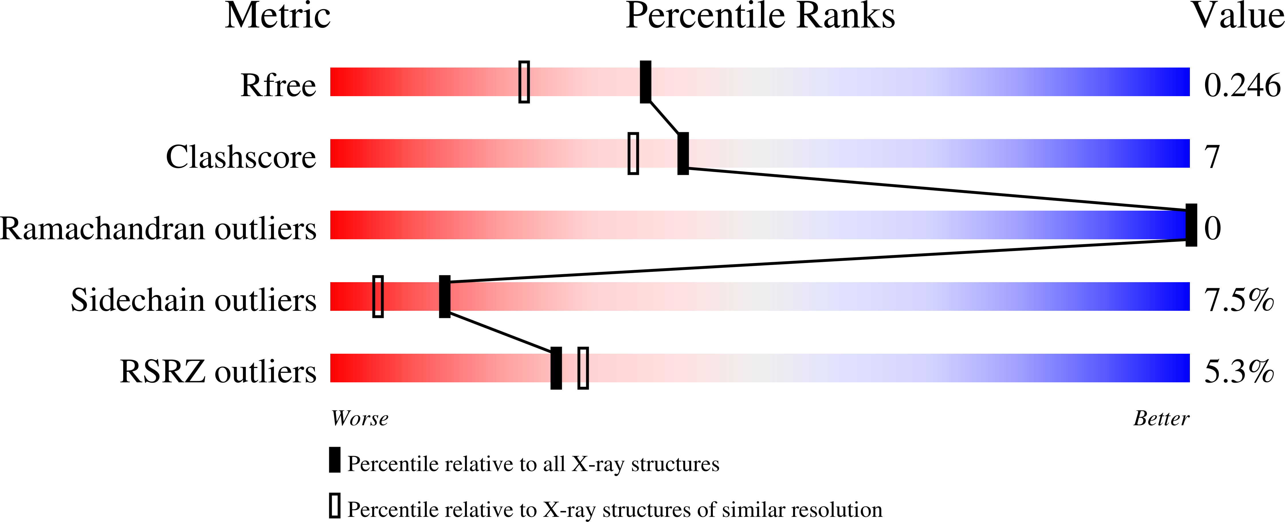
Resolution:
1.89 Å
R-Value Free:
0.24
R-Value Work:
0.20
R-Value Observed:
0.20
Space Group:
I 41 2 2
Macromolecular Entities
Structures with similar UniProt ID
Protein Blast
Polymer Type:polypeptide(L)
Molecule:Tetracycline repressor protein class D
Gene (Uniprot):tetR
Mutagens:A2S
Chain IDs:A
Chain Length:207
Number of Molecules:1
Biological Source:Escherichia coli
Primary Citation
Protonation Patterns in Tetracycline:Tet Repressor Recognition: Simulations and Experiments
Chembiochem 8 675 685 (2007)
PMID: 17361981 DOI: 10.1002/cbic.200600535

Abstact

Resistance to the antibiotic tetracycline (Tc) is regulated by its binding as a Tc:Mg2+ complex to the Tet Repressor protein (TetR). Tc:TetR recognition is a complex problem, with the protein and ligand each having several possible conformations and protonation states, which are difficult to elucidate by experiment alone. We used a combination of free-energy simulations and crystallographic analysis to investigate the electrostatic interactions between protein and ligand and the possible role of induced fit in Tc binding. Tc in solution was described quantum mechanically, while Tc:TetR interactions were described by a recent, high-quality molecular-mechanics model. The orientations of the amide and imidazole groups were determined experimentally by a careful analysis of Debye-Waller factors in alternate crystallographic models. The agreement with experiment for these orientations suggested that the simulations and their more detailed, thermodynamic predictions were reliable. We found that the ligand prefers an extended, zwitterionic state both in solution and in complexation with the protein. Tc is thus preorganized for binding, while the protein combines lock-and-key behavior for regions close to the ligand's amide, enolate, and ammonium groups, with an induced fit for regions close to the Mg2+ ion. These insights and the modeling techniques employed should be of interest for engineering improved TetR ligands and improved TetR proteins for gene regulation, as well as for drug design.

Legend

Protein

Chemical

Disease

Primary Citation of related structures
Feedback Form
Name
Email
Institute
Feedback