Deposition Date
2006-12-08
Release Date
2007-02-13
Last Version Date
2024-11-13
Entry Detail
PDB ID:
2O6V
Keywords:
Title:
Crystal structure and solution NMR studies of Lys48-linked tetraubiquitin at neutral pH
Biological Source:
Source Organism(s):
Homo sapiens (Taxon ID: 9606)
Expression System(s):
Method Details:
Experimental Method:
Resolution:
2.20 Å
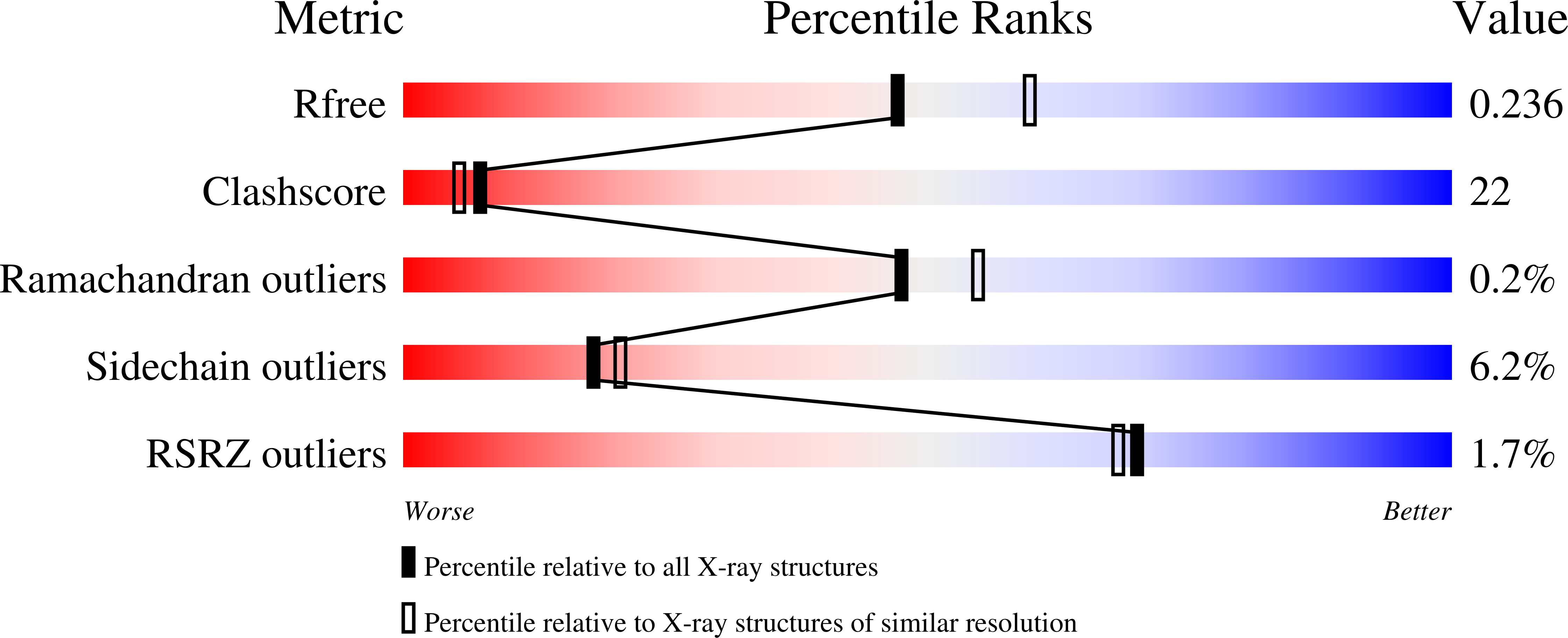
R-Value Free:
0.26
R-Value Work:
0.22
R-Value Observed:
0.24
Space Group:
C 1 2 1