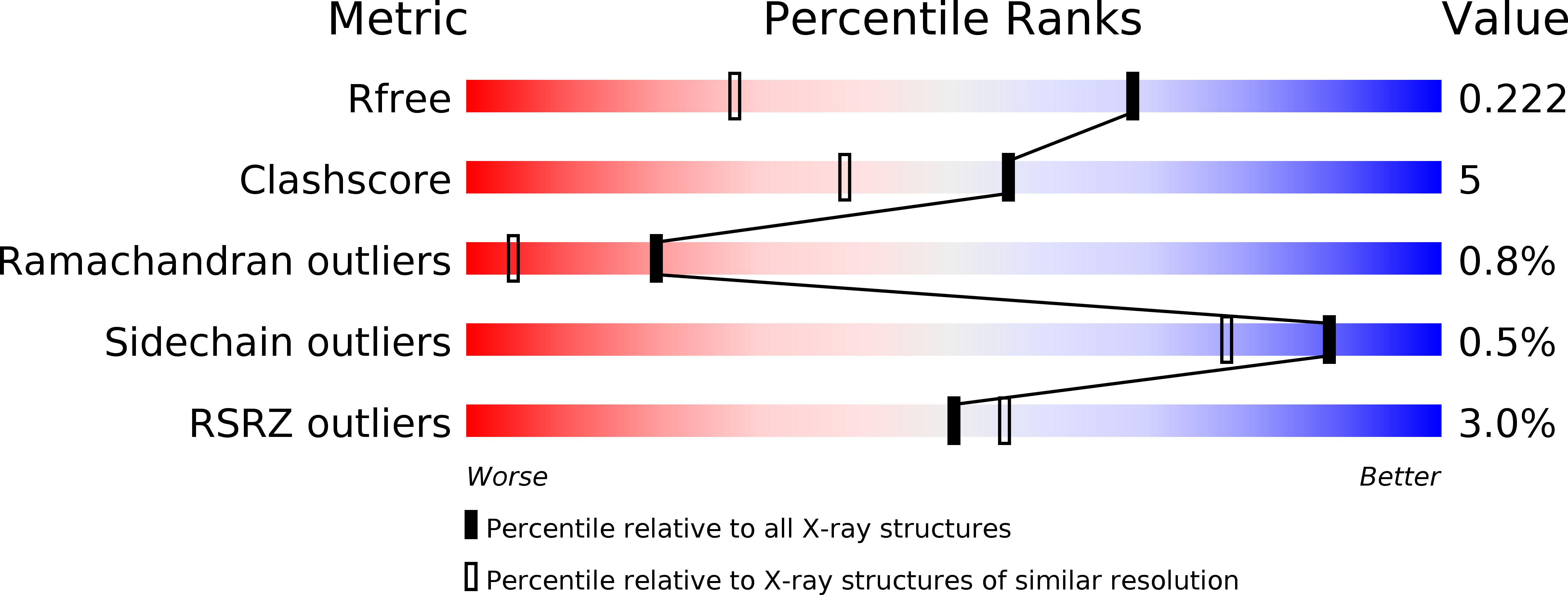
2O6P image
Deposition Date 2006-12-08
Release Date 2007-02-06
Last Version Date 2023-12-27
Entry Detail
PDB ID:
2O6P
Title:
Crystal Structure of the heme-IsdC complex
Biological Source:
Source Organism:
Host Organism:
Method Details:
Experimental Method:
Resolution:
1.50 Å
R-Value Free:
0.22
R-Value Work:
0.19
R-Value Observed:
0.19
Space Group:
P 21 21 21
Macromolecular Entities
Polymer Type:polypeptide(L)
Molecule:Iron-regulated surface determinant protein C
Chain IDs:A, B
Chain Length:161
Number of Molecules:2
Biological Source:Staphylococcus aureus subsp. aureus
Primary Citation
Crystal structure of the heme-IsdC complex, the central conduit of the Isd iron/heme uptake system in Staphylococcus aureus.
J. Biol. Chem. 282 10625 10631 (2007)
PMID: 17287214 DOI: 10.1074/jbc.M700234200

Abstact

Pathogens such as Staphylococcus aureus require iron to survive and have evolved specialized proteins to steal heme from their host. IsdC is the central conduit of the Isd (iron-regulated surface determinant) multicomponent heme uptake machinery; staphylococcal cell-surface proteins such as IsdA, IsdB, and IsdH are thought to funnel their molecular cargo to IsdC, which then mediates the transfer of the iron-containing nutrient to the membrane translocation system IsdDEF. The structure of the heme-IsdC complex reveals a novel heme site within an immunoglobulin-like domain and sheds light on its binding mechanism. The folding topology is reminiscent of the architecture of cytochrome f, cellobiose dehydrogenase, and ethylbenzene dehydrogenase; in these three proteins, the heme is bound in an equivalent position, but interestingly, IsdC features a distinct binding pocket with the ligand located next to the hydrophobic core of the beta-sandwich. The iron is coordinated with a tyrosine surrounded by several non-polar side chains that cluster into a tightly packed proximal side. On the other hand, the distal side is relatively exposed with a short helical peptide segment that acts as a lip clasping onto almost half of the porphyrin plane. This structural feature is argued to play a role in the mechanism of binding and release by switching to an open conformation and thus loosening the interactions holding the heme. The structure of the heme-IsdC complex provides a template for the understanding of other proteins, such as IsdA, IsdB, and IsdH, that contain the same heme-binding module as IsdC, known as the NEAT (near transporter) domain.

Legend

Protein

Chemical

Disease

Primary Citation of related structures