2O67 image
Deposition Date 2006-12-06
Release Date 2007-02-20
Last Version Date 2023-08-30
Entry Detail
PDB ID:
2O67
Title:
Crystal structure of Arabidopsis thaliana PII bound to malonate
Biological Source:
Source Organism:
Host Organism:
Method Details:
Experimental Method:
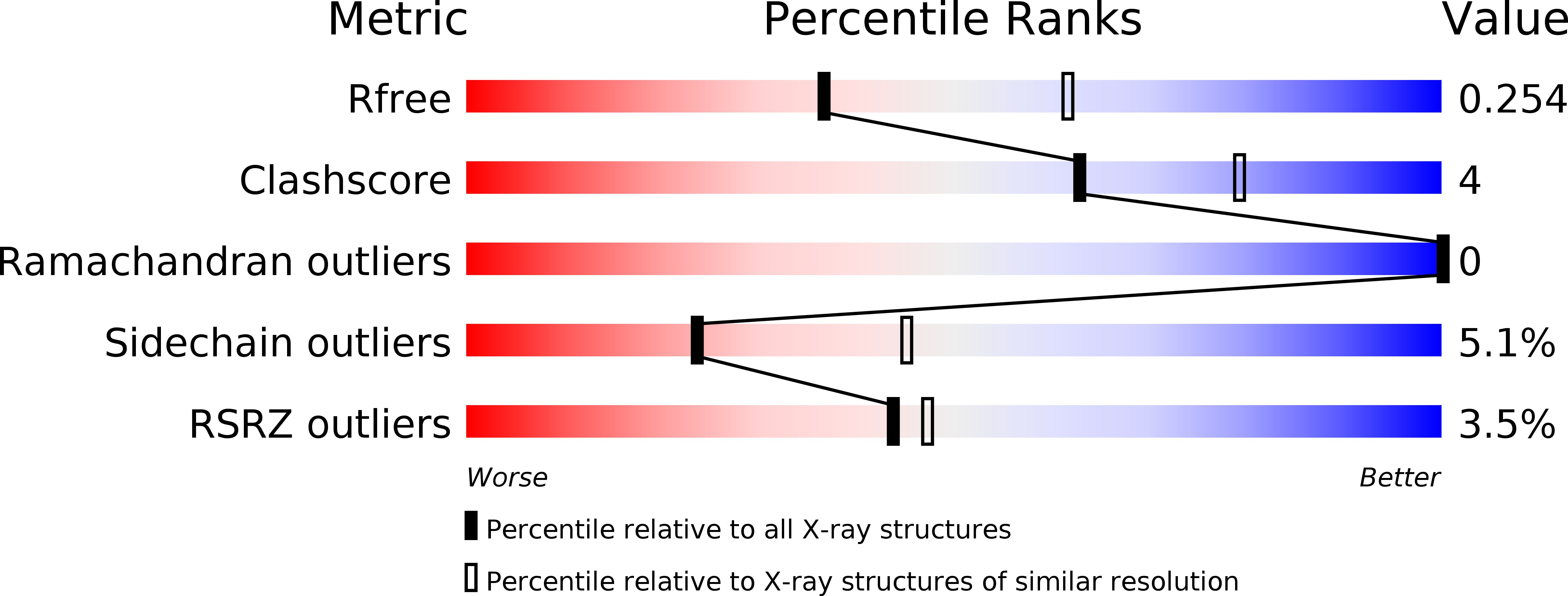
Resolution:
2.50 Å
R-Value Free:
0.26
R-Value Work:
0.20
R-Value Observed:
0.20
Space Group:
C 1 2 1
Macromolecular Entities
Structures with similar UniProt ID
Protein Blast
Polymer Type:polypeptide(L)
Molecule:PII protein
Gene (Uniprot):GLB1
Chain IDs:A, B, C
Chain Length:135
Number of Molecules:3
Biological Source:Arabidopsis thaliana
Ligand Molecules
Primary Citation
Crystal Structure of Arabidopsis PII Reveals Novel Structural Elements Unique to Plants.
Biochemistry 46 1477 1483 (2007)
PMID: 17279613 DOI: 10.1021/bi062149e

Abstact

The 1.9 A resolution crystal structure of PII from Arabidopsis thaliana reveals for the first time the molecular structure of a widely conserved regulator of carbon and nitrogen metabolism from a eukaryote. The structure provides a framework for understanding the arrangement of highly conserved residues shared with PII proteins from bacteria, archaea, and red algae as well as residues conserved only in plant PII. Most strikingly, a highly conserved segment at the N-terminus that is found only in plant PII forms numerous interactions with the alpha2 helix and projects from the surface of the homotrimer opposite to that occupied by the T-loop. In addition, solvent-exposed residues near the T-loop are highly conserved in plants but differ in prokaryotes. Several residues at the C-terminus that are also highly conserved only in plants contribute part of the ATP-binding site and likely participate in an ATP-induced conformational change. Structures of PII also reveal how citrate and malonate bind near the triphosphate binding site occupied by ATP in bacterial and archaeal PII proteins.

Legend

Protein

Chemical

Disease

Primary Citation of related structures