2O5E image
Entry Detail
PDB ID:
2O5E
Keywords:
Title:
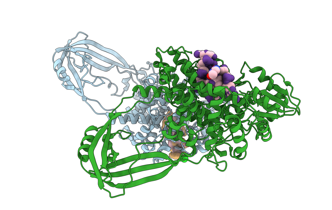
Structure of E. coli topoisomerase III in complex with an 8-base single stranded oligonucleotide. Frozen in glucose pH 7.0
Biological Source:
Source Organism:
Host Organism:
PDB Version:
Deposition Date:
2006-12-05
Release Date:
2007-04-03
Method Details:
Experimental Method:
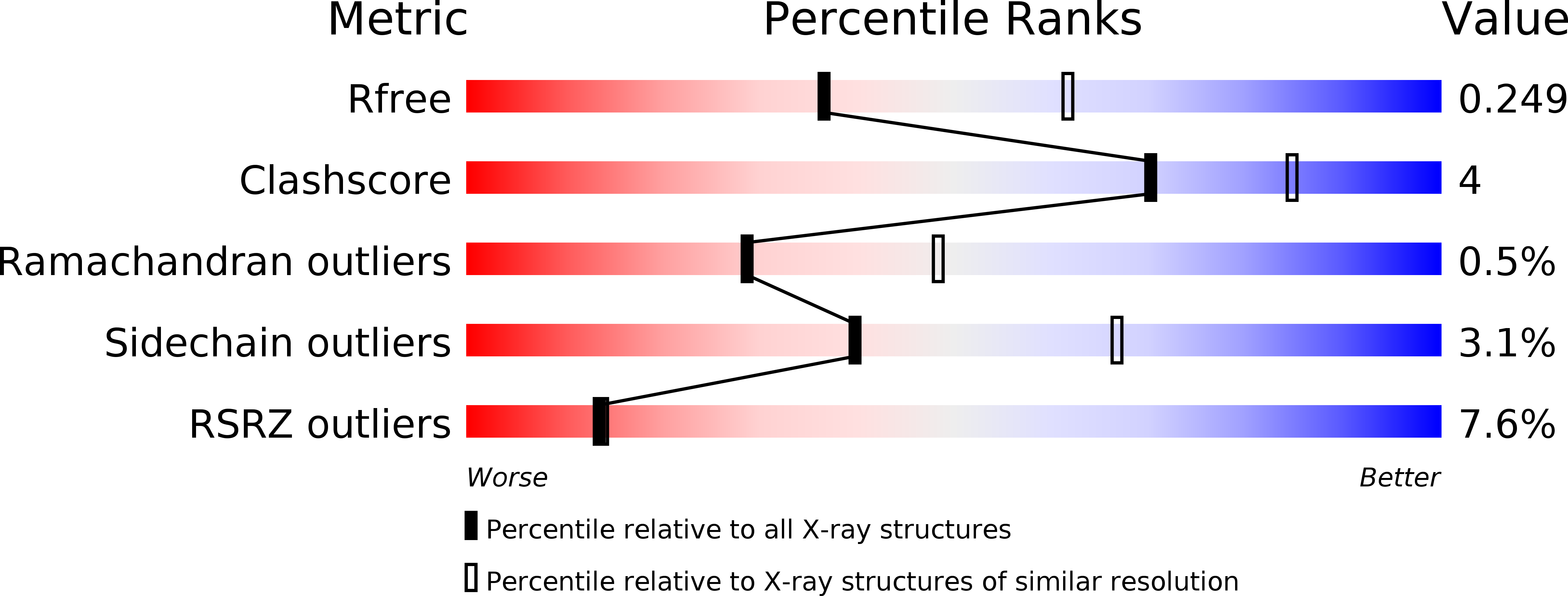
Resolution:
2.50 Å
R-Value Free:
0.26
R-Value Work:
0.21
R-Value Observed:
0.21
Space Group:
P 43 21 2
Macromolecular Entities
Polymer Type:polypeptide(L)
Description:DNA topoisomerase 3
Chain IDs:C (auth: A), D (auth: B)
Chain Length:659
Number of Molecules:2
Biological Source:Escherichia coli
Primary Citation
Structural Studies of E. coli Topoisomerase III-DNA Complexes Reveal a Novel Type IA Topoisomerase-DNA Conformational Intermediate.
J.Mol.Biol. 368 105 118 (2007)
PMID: 17331537 DOI: 10.1016/j.jmb.2007.01.065

Abstact

Escherichia coli DNA topoisomerase III belongs to the type IA family of DNA topoisomerases, which transiently cleave single-stranded DNA (ssDNA) via a 5' phosphotyrosine intermediate. We have solved crystal structures of wild-type E. coli topoisomerase III bound to an eight-base ssDNA molecule in three different pH environments. The structures reveal the enzyme in three distinct conformational states while bound to DNA. One conformation resembles the one observed previously with a DNA-bound, catalytically inactive mutant of topoisomerase III where DNA binding realigns catalytic residues to form a functional active site. Another conformation represents a novel intermediate in which DNA is bound along the ssDNA-binding groove but does not enter the active site, which remains in a catalytically inactive, closed state. A third conformation shows an intermediate state where the enzyme is still in a closed state, but the ssDNA is starting to invade the active site. For the first time, the active site region in the presence of both the catalytic tyrosine and ssDNA substrate is revealed for a type IA DNA topoisomerase, although there is no evidence of ssDNA cleavage. Comparative analysis of the various conformational states suggests a sequence of domain movements undertaken by the enzyme upon substrate binding.

Legend

Protein

Chemical

Disease

Primary Citation of related structures