2O2D image
Deposition Date 2006-11-29
Release Date 2007-11-13
Last Version Date 2023-08-30
Entry Detail
PDB ID:
2O2D
Keywords:
Title:
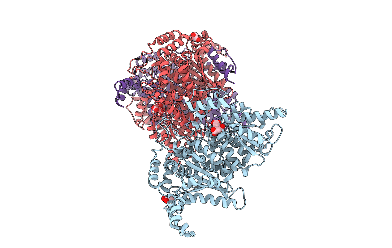
Crystal structure of phosphoglucose isomerase from Trypanosoma brucei complexed with citrate
Biological Source:
Source Organism:
Host Organism:
Method Details:
Experimental Method:
Resolution:
1.90 Å
R-Value Free:
0.23
R-Value Work:
0.21
Space Group:
C 2 2 21
Macromolecular Entities
Polymer Type:polypeptide(L)
Molecule:Glucose-6-phosphate isomerase, glycosomal
Gene (Uniprot):PGI
Chain IDs:A, B, C
Chain Length:613
Number of Molecules:3
Biological Source:Trypanosoma brucei brucei
Primary Citation
Crystal structure of phosphoglucose isomerase from Trypanosoma brucei complexed with glucose-6-phosphate at 1.6 A resolution
Proteins 74 72 80 (2008)
PMID: 18561188 DOI: 10.1002/prot.22133

Abstact

Enzymes of glycolysis in Trypanosoma brucei have been identified as potential drug targets for African sleeping sickness because glycolysis is the only source of ATP for the bloodstream form of this parasite. Several inhibitors were previously reported to bind preferentially to trypanosomal phosphoglucose isomerase (PGI, the second enzyme in glycolysis) than to mammalian PGIs, which suggests that PGI might make a good target for species-specific drug design. Herein, we report recombinant expression, purification, crystallization and X-ray crystal structure determination of T. brucei PGI. One structure solved at 1.6 A resolution contains a substrate, D-glucose-6-phosphate, in an extended conformation in the active site. A second structure solved at 1.9 A resolution contains a citrate molecule in the active site. The structures are compared with the crystal structures of PGI from humans and from Leishmania mexicana. The availability of recombinant tPGI and its first high-resolution crystal structures are initial steps in considering this enzyme as a potential drug target.

Legend

Protein

Chemical

Disease

Primary Citation of related structures