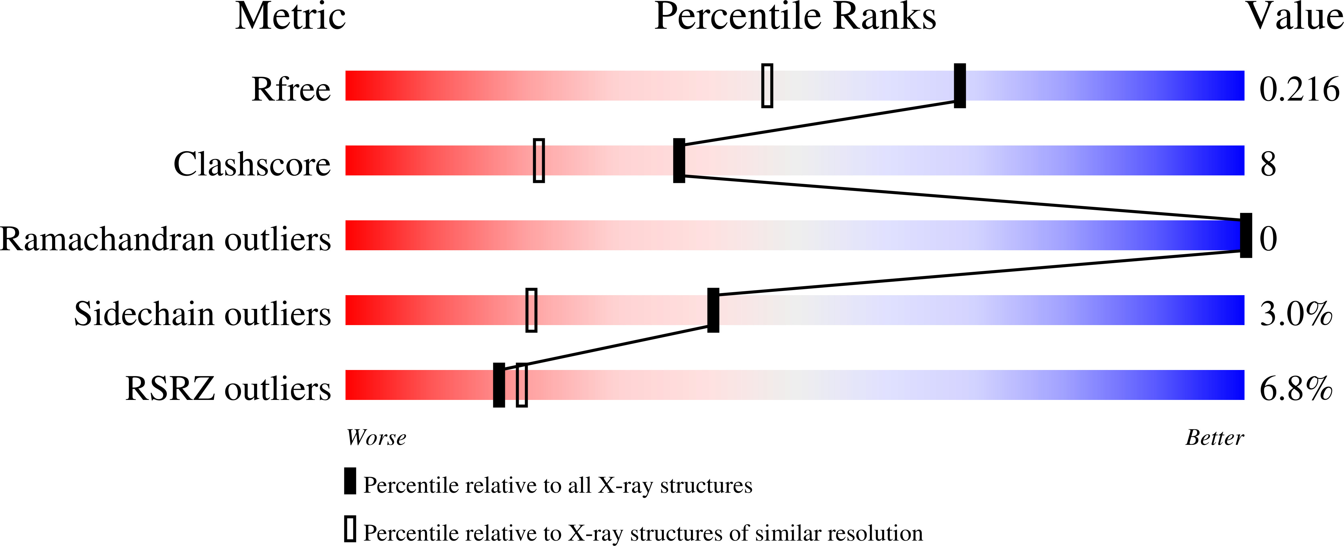
2O1G image
Deposition Date 2006-11-28
Release Date 2007-02-06
Last Version Date 2023-08-30
Entry Detail
PDB ID:
2O1G
Keywords:
Title:
Natural occurring mutant of Human ABO(H) Galactosyltransferase: GTB/M214T
Biological Source:
Source Organism:
Homo sapiens (Taxon ID: 9606)
Host Organism:
Method Details:
Experimental Method:
Resolution:
1.71 Å
R-Value Free:
0.21
R-Value Work:
0.18
R-Value Observed:
0.18
Space Group:
C 2 2 21
Macromolecular Entities
Structures with similar UniProt ID
Protein Blast
Polymer Type:polypeptide(L)
Molecule:ABO glycosyltransferase
Chain IDs:A
Chain Length:297
Number of Molecules:1
Biological Source:Homo sapiens
Ligand Molecules
Primary Citation
Structural effects of naturally occurring human blood group B galactosyltransferase mutations adjacent to the DXD motif.
J.Biol.Chem. 282 9564 9570 (2007)
PMID: 17259183 DOI: 10.1074/jbc.M610998200

Abstact

Human blood group A and B antigens are produced by two closely related glycosyltransferase enzymes. An N-acetylgalactosaminyltransferase (GTA) utilizes UDP-GalNAc to extend H antigen acceptors (Fuc alpha(1-2)Gal beta-OR) producing A antigens, whereas a galactosyltransferase (GTB) utilizes UDP-Gal as a donor to extend H structures producing B antigens. GTA and GTB have a characteristic (211)DVD(213) motif that coordinates to a Mn(2+) ion shown to be critical in donor binding and catalysis. Three GTB mutants, M214V, M214T, and M214R, with alterations adjacent to the (211)DVD(213) motif have been identified in blood banking laboratories. From serological phenotyping, individuals with the M214R mutation show the B(el) variant expressing very low levels of B antigens, whereas those with M214T and M214V mutations give rise to A(weak)B phenotypes. Kinetic analysis of recombinant mutant GTB enzymes revealed that M214R has a 1200-fold decrease in k(cat) compared with wild type GTB. The crystal structure of M214R showed that DVD motif coordination to Mn(2+) was disrupted by Arg-214 causing displacement of the metal by a water molecule. Kinetic characterizations of the M214T and M214V mutants revealed they both had GTA and GTB activity consistent with the serology. The crystal structure of the M214T mutant showed no change in DVD coordination to Mn(2+). Instead a critical residue, Met-266, which is responsible for determining donor specificity, had adopted alternate conformations. The conformation with the highest occupancy opens up the active site to accommodate the larger A-specific donor, UDP-GalNAc, accounting for the dual specificity.

Legend

Protein

Chemical

Disease

Primary Citation of related structures