
Deposition Date
2006-11-20
Release Date
2006-12-05
Last Version Date
2023-08-30
Entry Detail
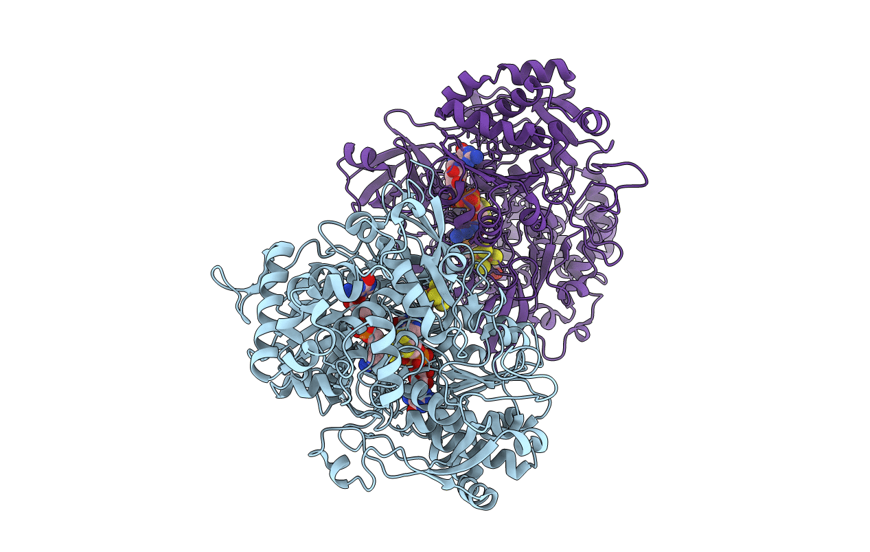
PDB ID:
2NYA
Keywords:
Title:
Crystal structure of the periplasmic nitrate reductase (NAP) from Escherichia coli
Biological Source:
Source Organism:
Escherichia coli K12 (Taxon ID: 83333)
Host Organism:
Method Details:
Experimental Method:
Resolution:
2.50 Å
R-Value Free:
0.24
R-Value Work:
0.18
R-Value Observed:
0.18
Space Group:
P 1 21 1
