2NXT image
Deposition Date 2006-11-19
Release Date 2007-04-24
Last Version Date 2023-08-30
Entry Detail
PDB ID:
2NXT
Keywords:
Title:
Structural and kinetic effects of hydrophobic mutations in the active site of human carbonic anhydrase II
Biological Source:
Source Organism:
Homo sapiens (Taxon ID: 9606)
Host Organism:
Method Details:
Experimental Method:
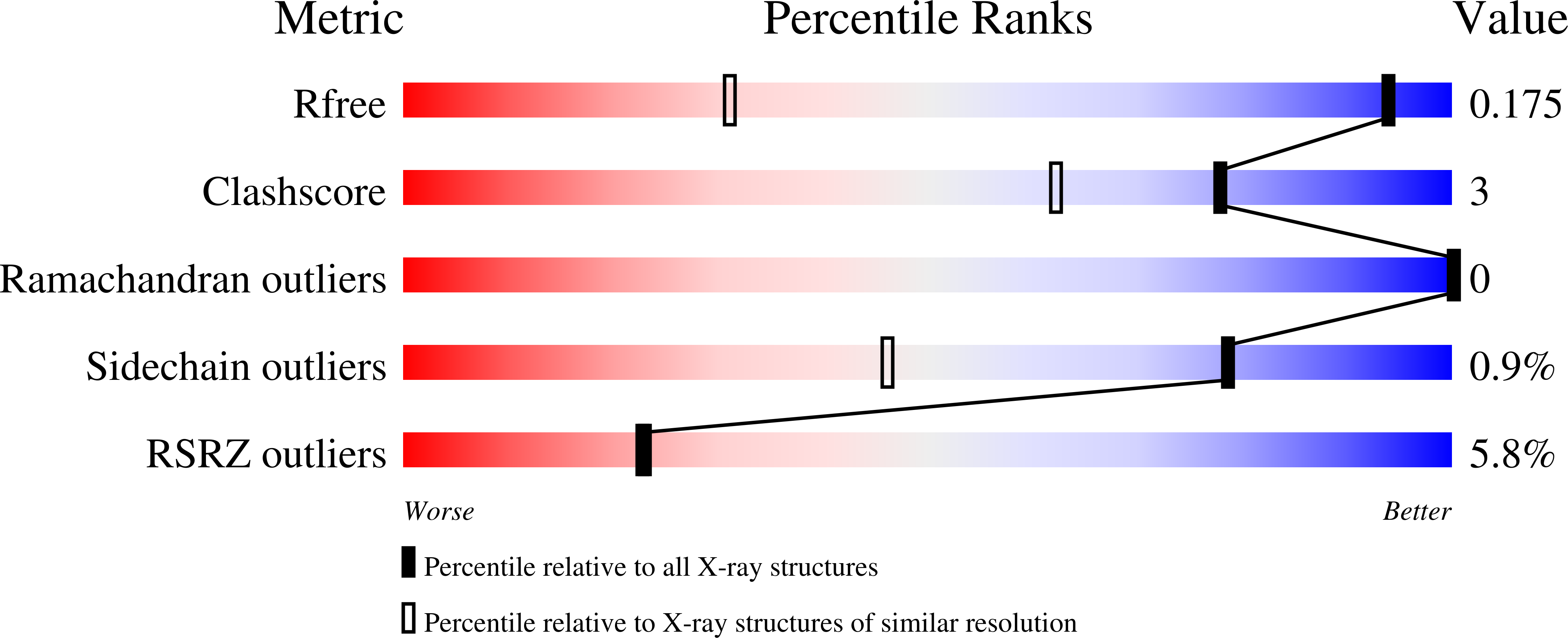
Resolution:
1.15 Å
R-Value Free:
0.18
R-Value Work:
0.16
R-Value Observed:
0.16
Space Group:
P 1 21 1
Macromolecular Entities
Polymer Type:polypeptide(L)
Molecule:carbonic anhydrase 2
Gene (Uniprot):CA2
Mutations:Y7F
Chain IDs:A
Chain Length:260
Number of Molecules:1
Biological Source:Homo sapiens
Ligand Molecules
Primary Citation
Speeding Up Proton Transfer in a Fast Enzyme: Kinetic and Crystallographic Studies on the Effect of Hydrophobic Amino Acid Substitutions in the Active Site of Human Carbonic Anhydrase II.
Biochemistry 46 3803 3813 (2007)
PMID: 17330962 DOI: 10.1021/bi602620k

Abstact

Catalysis of the hydration of CO2 by human carbonic anhydrase isozyme II (HCA II) is sustained at a maximal catalytic turnover of 1 mus-1 by proton transfer between a zinc-bound solvent and bulk solution. This mechanism of proton transfer is facilitated via the side chain of His64, which is located 7.5 A from the zinc, and mediated via intervening water molecules in the active-site cavity. Three hydrophilic residues that have previously been shown to contribute to the stabilization of these intervening waters were replaced with hydrophobic residues (Y7F, N62L, and N67L) to determine their effects on proton transfer. The structures of all three mutants were determined by X-ray crystallography, with crystals equilibrated from pH 6.0 to 10.0. A range of changes were observed in the ordered solvent and the conformation of the side chain of His64. Correlating these structural variants with kinetic studies suggests that the very efficient proton transfer (approximately 7 micros-1) observed for Y7F HCA II in the dehydration direction, compared with the wild type and other mutants of this study, is due to a combination of three features. First, in this mutant, the side chain of His64 showed an appreciable inward orientation pointing toward the active-site zinc. Second, in the structure of Y7F HCA II, there is an unbranched chain of hydrogen-bonded waters linking the proton donor His64 and acceptor zinc-bound hydroxide. Finally, the difference in pKa of the donor and acceptor appears favorable for proton transfer. The data suggest roles for residues 7, 62, and 67 in fine-tuning the properties of His64 for optimal proton transfer in catalysis.

Legend

Protein

Chemical

Disease

Primary Citation of related structures