2NWG image
Deposition Date 2006-11-14
Release Date 2007-02-13
Last Version Date 2024-10-30
Entry Detail
PDB ID:
2NWG
Title:
Structure of CXCL12:heparin disaccharide complex
Biological Source:
Source Organism:
Homo sapiens (Taxon ID: 9606)
Method Details:
Experimental Method:
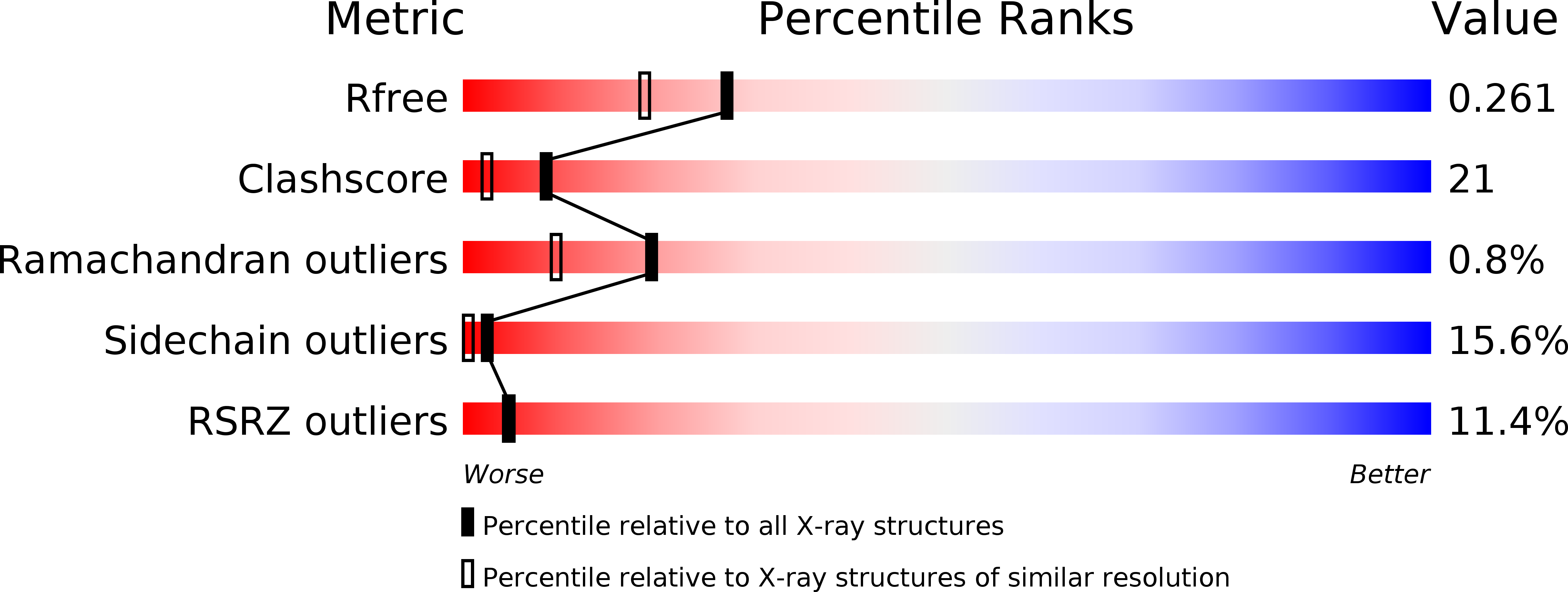
Resolution:
2.07 Å
R-Value Free:
0.26
R-Value Work:
0.24
R-Value Observed:
0.24
Space Group:
P 21 21 21
Macromolecular Entities
Structures with similar UniProt ID
Protein Blast
Polymer Type:polypeptide(L)
Molecule:Stromal cell-derived factor 1
Gene (Uniprot):CXCL12
Chain IDs:A, B
Chain Length:68
Number of Molecules:2
Biological Source:Homo sapiens
Peptide-like Molecules
PRD_900026
Primary Citation
Structural and Functional Basis of CXCL12 (Stromal Cell-derived Factor-1{alpha}) Binding to Heparin
J.Biol.Chem. 282 10018 10027 (2007)
PMID: 17264079 DOI: 10.1074/jbc.M608796200

Abstact

CXCL12 (SDF-1alpha) and CXCR4 are critical for embryonic development and cellular migration in adults. These proteins are involved in HIV-1 infection, cancer metastasis, and WHIM disease. Sequestration and presentation of CXCL12 to CXCR4 by glycosaminoglycans (GAGs) is proposed to be important for receptor activation. Mutagenesis has identified CXCL12 residues that bind to heparin. However, the molecular details of this interaction have not yet been determined. Here we demonstrate that soluble heparin and heparan sulfate negatively affect CXCL12-mediated in vitro chemotaxis. We also show that a cluster of basic residues in the dimer interface is required for chemotaxis and is a target for inhibition by heparin. We present structural evidence for binding of an unsaturated heparin disaccharide to CXCL12 attained through solution NMR spectroscopy and x-ray crystallography. Increasing concentrations of the disaccharide altered the two-dimensional (1)H-(15)N-HSQC spectra of CXCL12, which identified two clusters of residues. One cluster corresponds to beta-strands in the dimer interface. The second includes the amino-terminal loop and the alpha-helix. In the x-ray structure two unsaturated disaccharides are present. One is in the dimer interface with direct contacts between residues His(25), Lys(27), and Arg(41) of CXCL12 and the heparin disaccharide. The second disaccharide contacts Ala(20), Arg(21), Asn(30), and Lys(64). This is the first x-ray structure of a CXC class chemokine in complex with glycosaminoglycans. Based on the observation of two heparin binding sites, we propose a mechanism in which GAGs bind around CXCL12 dimers as they sequester and present CXCL12 to CXCR4.

Legend

Protein

Chemical

Disease

Primary Citation of related structures