Deposition Date
2006-11-09
Release Date
2007-11-20
Last Version Date
2023-08-30
Entry Detail
PDB ID:
2NUG
Keywords:
Title:
Crystal structure of RNase III from Aquifex aeolicus complexed with ds-RNA at 1.7-Angstrom Resolution
Biological Source:
Source Organism(s):
Aquifex aeolicus (Taxon ID: 63363)
Expression System(s):
Method Details:
Experimental Method:
Resolution:
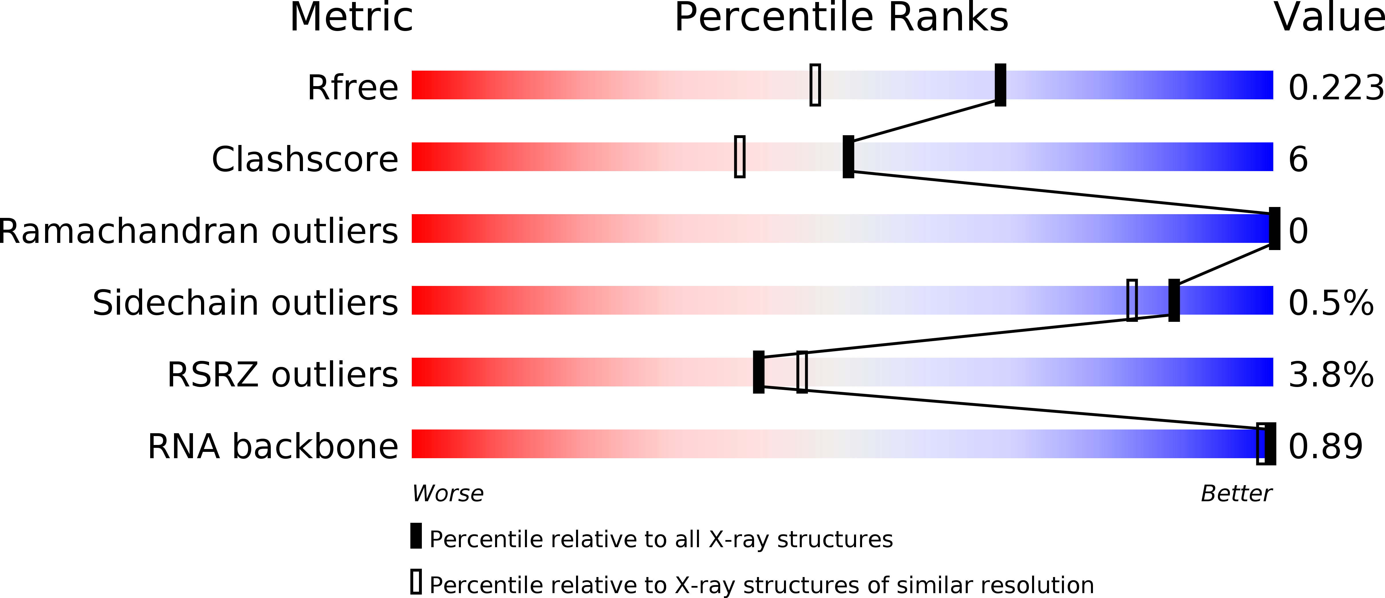
1.70 Å
R-Value Free:
0.22
R-Value Work:
0.19
R-Value Observed:
0.19
Space Group:
P 21 21 2