2NU5 image
Deposition Date 2006-11-08
Release Date 2007-08-07
Last Version Date 2024-10-30
Entry Detail
PDB ID:
2NU5
Title:
Crystal structure of a complex of griffithsin cocrystallized with N-acetylglucosamine
Biological Source:
Source Organism:
Host Organism:
Method Details:
Experimental Method:
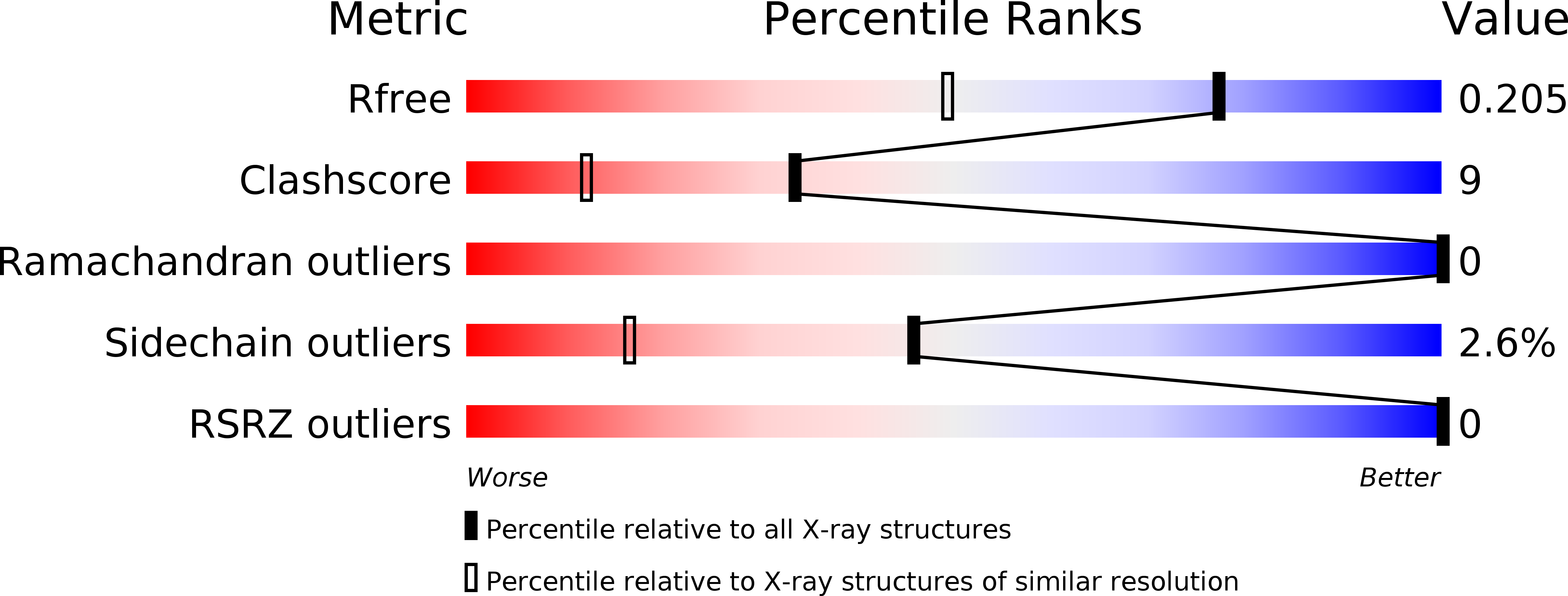
Resolution:
1.56 Å
R-Value Free:
0.20
R-Value Work:
0.16
R-Value Observed:
0.16
Space Group:
P 21 21 21
Macromolecular Entities
Polymer Type:polypeptide(L)
Molecule:griffithsin
Chain IDs:A, B
Chain Length:122
Number of Molecules:2
Biological Source:Griffithsia sp.
Primary Citation
Crystallographic studies of the complexes of antiviral protein griffithsin with glucose and N-acetylglucosamine
Protein Sci. 16 1485 1489 (2007)
PMID: 17567736 DOI: 10.1110/ps.072889407

Abstact

Crystal structures of complexes of an antiviral lectin griffithsin (GRFT) with glucose and N-acetylglucosamine were solved and refined at high resolution. In both complexes, all six monosaccharide-binding sites of GRFT were occupied and the mode of binding was similar to that of mannose. In our previous attempts to obtain a complex with N-acetylglucosamine by soaking, only a single site was occupied; thus, cocrystallization was clearly superior despite lower concentration of the ligand. Isothermal titration calorimetric experiments with N-acetylglucosamine, glucose, and mannose provided enthalpic evidence of distinct binding differences between the three monosaccharides. A comparison of the mode of binding of different monosaccharides is discussed in the context of the antiviral activity of GRFT, based on specific binding to high-mannose-containing complex carbohydrates found on viral envelopes.

Legend

Protein

Chemical

Disease

Primary Citation of related structures