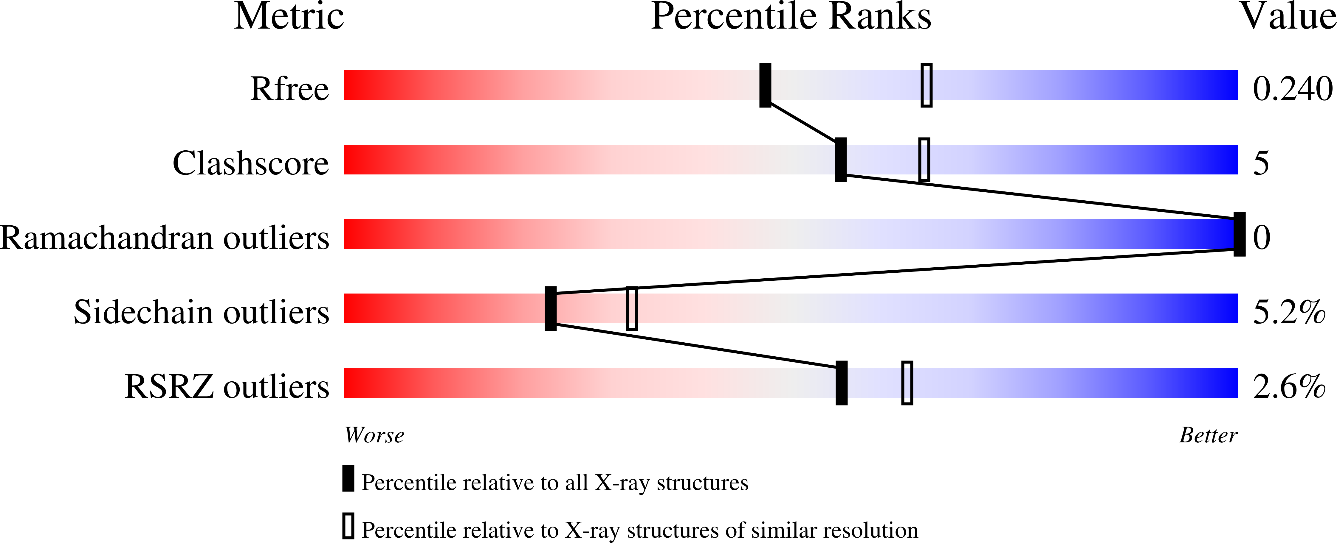
2NTN image
Deposition Date 2006-11-08
Release Date 2006-11-21
Last Version Date 2023-10-25
Entry Detail
PDB ID:
2NTN
Keywords:
Title:
Crystal structure of MabA-C60V/G139A/S144L
Biological Source:
Source Organism:
Method Details:
Experimental Method:
Resolution:
2.30 Å
R-Value Free:
0.23
R-Value Work:
0.17
R-Value Observed:
0.18
Space Group:
C 1 2 1
Macromolecular Entities
Polymer Type:polypeptide(L)
Molecule:3-oxoacyl-[acyl-carrier-protein] reductase
Mutations:C60V, G139A, S144L
Chain IDs:A, B
Chain Length:267
Number of Molecules:2
Biological Source:Mycobacterium tuberculosis H37Rv
Primary Citation
Lack of dynamics in the MabA active site kills the enzyme activity: practical consequences for drug-design studies
ACTA CRYSTALLOGR.,SECT.D 63 923 925 (2007)
PMID: 17642518 DOI: 10.1107/S0907444907024158

Abstact

The MabA protein from Mycobacterium tuberculosis is a validated drug target. Previous structural studies of this protein showed dynamic behaviour in the catalytic site and described motion between an open 'active' holo form (with NADP) and a closed 'inactive' apo form (without NADP). Here, a mutation (G139A) is reported that leads to complete protein inactivation and freezes the catalytic site into its closed form, even in the presence of the cofactor. This observation suggests a new way to develop anti-MabA drugs via protein stabilization of the 'inactive' form.

Legend

Protein

Chemical

Disease

Primary Citation of related structures