2NSP image
Deposition Date 2006-11-06
Release Date 2007-09-18
Last Version Date 2024-10-30
Entry Detail
PDB ID:
2NSP
Keywords:
Title:
Crystal structure of pectin methylesterase D178A mutant in complex with hexasaccharide I
Biological Source:
Source Organism:
Host Organism:
Method Details:
Experimental Method:
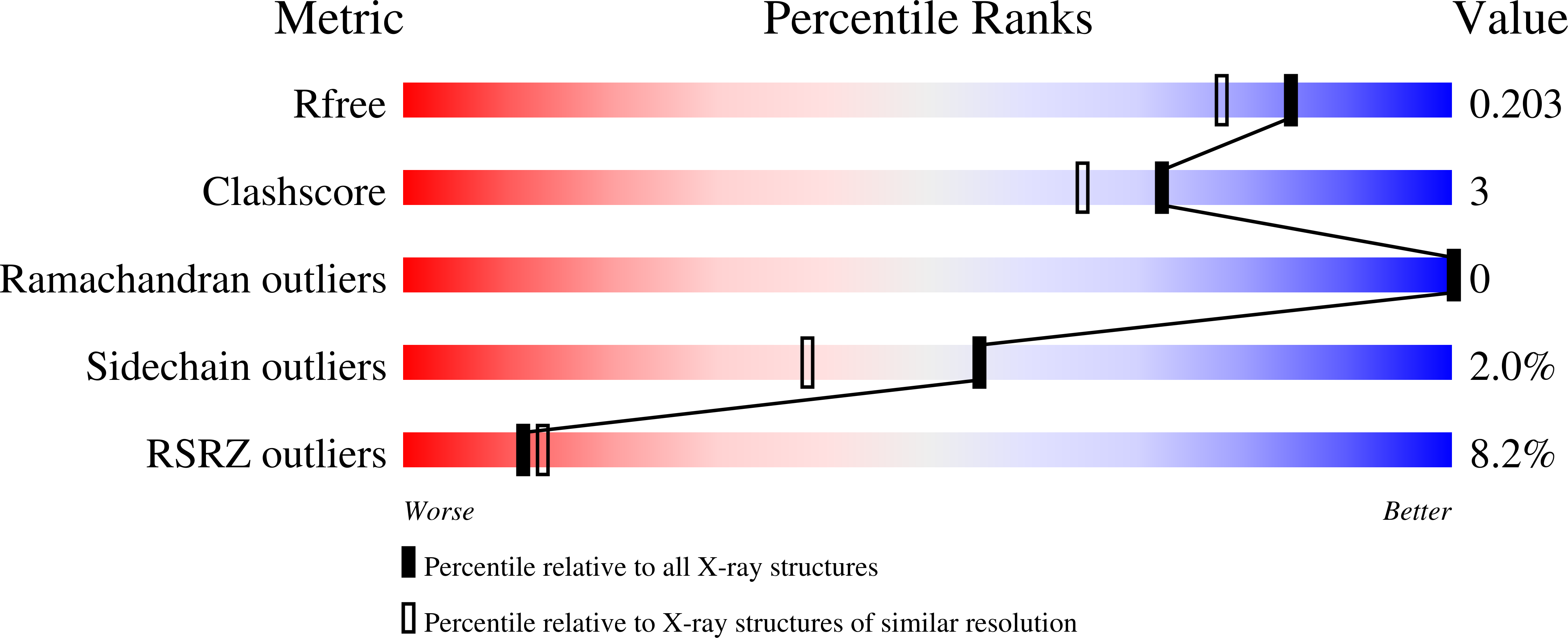
Resolution:
1.70 Å
R-Value Free:
0.20
R-Value Work:
0.18
R-Value Observed:
0.18
Space Group:
P 1 21 1
Macromolecular Entities
Polymer Type:polypeptide(L)
Molecule:Pectinesterase A
Gene (Uniprot):pemA
Mutations:D178A
Chain IDs:A, B
Chain Length:342
Number of Molecules:2
Biological Source:Erwinia chrysanthemi
Primary Citation
Molecular basis of the activity of the phytopathogen pectin methylesterase.
Embo J. 26 3879 3887 (2007)
PMID: 17717531 DOI: 10.1038/sj.emboj.7601816

Abstact

We provide a mechanism for the activity of pectin methylesterase (PME), the enzyme that catalyses the essential first step in bacterial invasion of plant tissues. The complexes formed in the crystal using specifically methylated pectins, together with kinetic measurements of directed mutants, provide clear insights at atomic resolution into the specificity and the processive action of the Erwinia chrysanthemi enzyme. Product complexes provide additional snapshots along the reaction coordinate. We previously revealed that PME is a novel aspartic-esterase possessing parallel beta-helix architecture and now show that the two conserved aspartates are the nucleophile and general acid-base in the mechanism, respectively. Other conserved residues at the catalytic centre are shown to be essential for substrate binding or transition state stabilisation. The preferential binding of methylated sugar residues upstream of the catalytic site, and demethylated residues downstream, drives the enzyme along the pectin molecule and accounts for the sequential pattern of demethylation produced by both bacterial and plant PMEs.

Legend

Protein

Chemical

Disease

Primary Citation of related structures