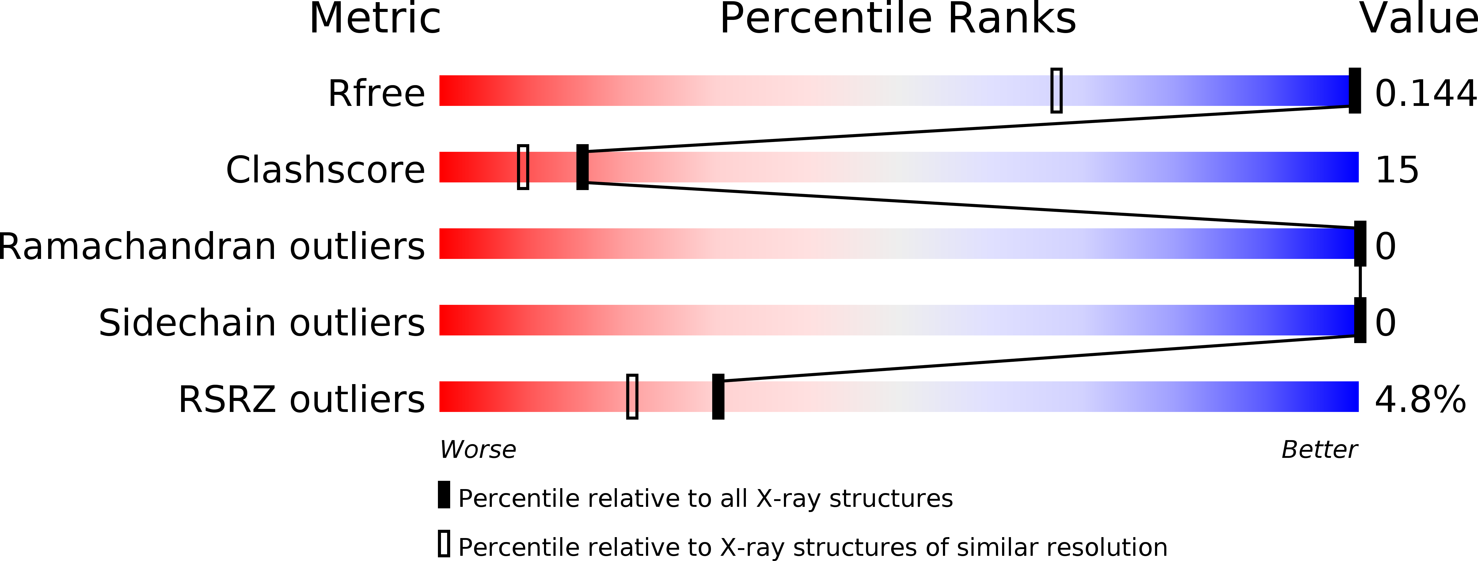
2NRL image
Deposition Date 2006-11-02
Release Date 2007-05-08
Last Version Date 2024-11-06
Entry Detail
PDB ID:
2NRL
Title:
Blackfin tuna myoglobin
Biological Source:
Source Organism:
Method Details:
Experimental Method:
Resolution:
0.91 Å
R-Value Free:
0.14
R-Value Work:
0.14
R-Value Observed:
0.14
Space Group:
P 1 21 1
Macromolecular Entities
Polymer Type:polypeptide(L)
Molecule:Myoglobin
Chain IDs:A
Chain Length:147
Number of Molecules:1
Biological Source:Thunnus atlanticus
Primary Citation
S-nitrosylation-induced conformational change in blackfin tuna myoglobin.
J.Biol.Chem. 282 19773 19780 (2007)
PMID: 17488722 DOI: 10.1074/jbc.M701363200

Abstact

S-nitrosylation is a post-translational protein modification that can alter the function of a variety of proteins. Despite the growing wealth of information that this modification may have important functional consequences, little is known about the structure of the moiety or its effect on protein tertiary structure. Here we report high-resolution x-ray crystal structures of S-nitrosylated and unmodified blackfin tuna myoglobin, which demonstrate that in vitro S-nitrosylation of this protein at the surface-exposed Cys-10 directly causes a reversible conformational change by "wedging" apart a helix and loop. Furthermore, we have demonstrated in solution and in a single crystal that reduction of the S-nitrosylated myoglobin with dithionite results in NO cleavage from the sulfur of Cys-10 and rebinding to the reduced heme iron, showing the reversibility of both the modification and the conformational changes. Finally, we report the 0.95-A structure of ferrous nitrosyl myoglobin, which provides an accurate structural view of the NO coordination geometry in the context of a globin heme pocket.

Legend

Protein

Chemical

Disease

Primary Citation of related structures