2NRF image
Deposition Date 2006-11-02
Release Date 2006-11-14
Last Version Date 2023-08-30
Entry Detail
PDB ID:
2NRF
Title:
Crystal Structure of GlpG, a Rhomboid family intramembrane protease
Biological Source:
Source Organism:
Escherichia coli (Taxon ID: 562)
Host Organism:
Method Details:
Experimental Method:
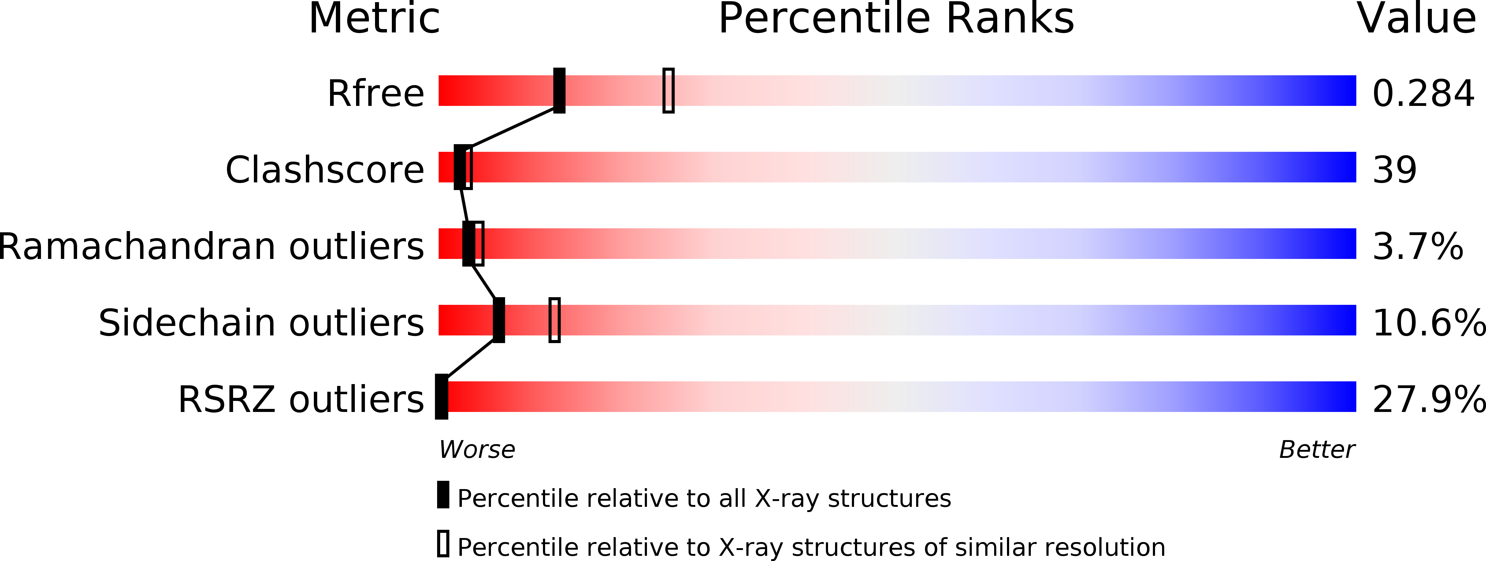
Resolution:
2.60 Å
R-Value Free:
0.29
R-Value Work:
0.26
R-Value Observed:
0.28
Space Group:
P 31
Macromolecular Entities
Structures with similar UniProt ID
Protein Blast
Polymer Type:polypeptide(L)
Molecule:Protein GlpG
Gene (Uniprot):glpG
Chain IDs:A, B
Chain Length:182
Number of Molecules:2
Biological Source:Escherichia coli
Primary Citation
Structural analysis of a rhomboid family intramembrane protease reveals a gating mechanism for substrate entry.
Nat.Struct.Mol.Biol. 13 1084 1091 (2006)
PMID: 17099694 DOI: 10.1038/nsmb1179

Abstact

Intramembrane proteolysis regulates diverse biological processes. Cleavage of substrate peptide bonds within the membrane bilayer is catalyzed by integral membrane proteases. Here we report the crystal structure of the transmembrane core domain of GlpG, a rhomboid-family intramembrane serine protease from Escherichia coli. The protein contains six transmembrane helices, with the catalytic Ser201 located at the N terminus of helix alpha4 approximately 10 A below the membrane surface. Access to water molecules is provided by a central cavity that opens to the extracellular region and converges on Ser201. One of the two GlpG molecules in the asymmetric unit has an open conformation at the active site, with the transmembrane helix alpha5 bent away from the rest of the molecule. Structural analysis suggests that substrate entry to the active site is probably gated by the movement of helix alpha5.

Legend

Protein

Chemical

Disease

Primary Citation of related structures