2NRB image
Deposition Date 2006-11-01
Release Date 2007-09-18
Last Version Date 2023-08-30
Entry Detail
PDB ID:
2NRB
Keywords:
Title:
C28S Mutant of Succinyl-CoA:3-Ketoacid CoA Transferase from Pig Heart
Biological Source:
Source Organism:
Sus scrofa (Taxon ID: 9823)
Method Details:
Experimental Method:
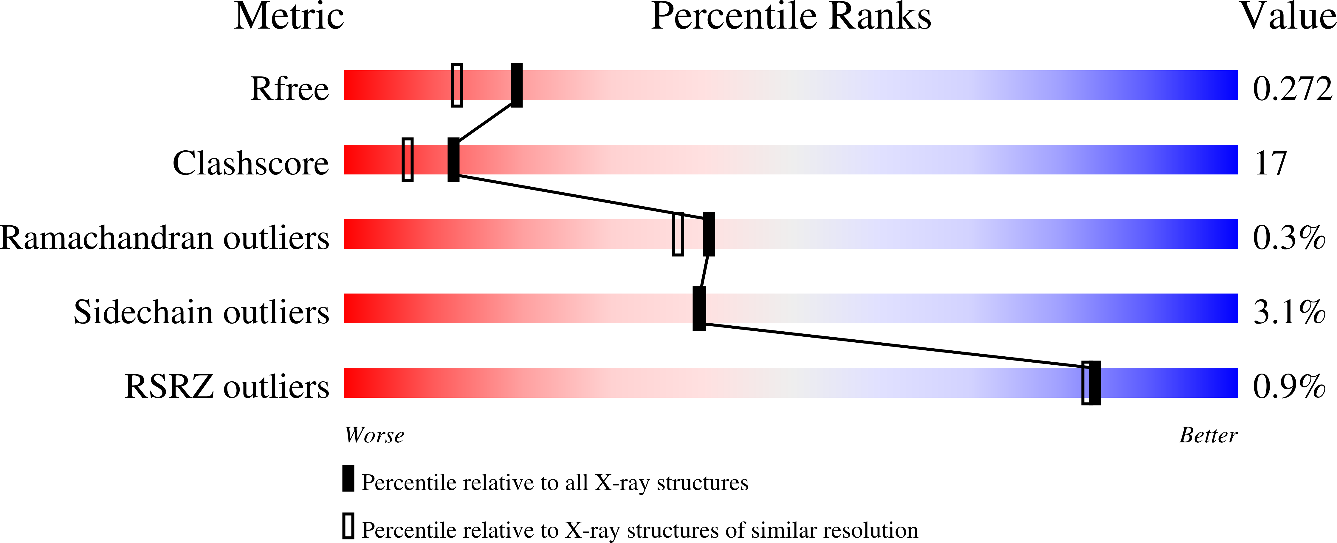
Resolution:
2.00 Å
R-Value Free:
0.27
R-Value Work:
0.22
R-Value Observed:
0.22
Space Group:
P 1 21 1
Macromolecular Entities
Structures with similar UniProt ID
Protein Blast
Polymer Type:polypeptide(L)
Molecule:Succinyl-CoA:3-ketoacid-coenzyme A transferase 1
Gene (Uniprot):OXCT1
Mutagens:C28S
Chain IDs:A, B, C, D
Chain Length:481
Number of Molecules:4
Biological Source:Sus scrofa
Ligand Molecules
Primary Citation
Identification of the Cysteine Residue Exposed by the Conformational Change in Pig Heart Succinyl-CoA:3-Ketoacid Coenzyme A Transferase on Binding Coenzyme A.
Biochemistry 46 10852 10863 (2007)
PMID: 17718512 DOI: 10.1021/bi700828h

Abstact

Succinyl-CoA:3-ketoacid CoA transferase (SCOT) transfers CoA from succinyl-CoA to acetoacetate via a thioester intermediate with its active site glutamate residue, Glu 305. When CoA is linked to the enzyme, a cysteine residue can now be rapidly modified by 5,5'-dithiobis(2-nitrobenzoic acid), reflecting a conformational change of SCOT upon formation of the thioester. Since either Cys 28 or Cys 196 could be the target, each was mutated to Ser to distinguish between them. Like wild-type SCOT, the C196S mutant protein was modified rapidly in the presence of acyl-CoA substrates. In contrast, the C28S mutant protein was modified much more slowly under identical conditions, indicating that Cys 28 is the residue exposed on binding CoA. The specific activity of the C28S mutant protein was unexpectedly lower than that of wild-type SCOT. X-ray crystallography revealed that Ser adopts a different conformation than the native Cys. A chloride ion is bound to one of four active sites in the crystal structure of the C28S mutant protein, mimicking substrate, interacting with Lys 329, Asn 51, and Asn 52. On the basis of these results and the studies of the structurally similar CoA transferase from Escherichia coli, YdiF, bound to CoA, the conformational change in SCOT was deduced to be a domain rotation of 17 degrees coupled with movement of two loops: residues 321-329 that bury Cys 28 and interact with succinate or acetoacetate and residues 374-386 that interact with CoA. Modeling this conformational change has led to the proposal of a new mechanism for catalysis by SCOT.

Legend

Protein

Chemical

Disease

Primary Citation of related structures