2NR6 image
Deposition Date 2006-11-01
Release Date 2008-02-19
Last Version Date 2023-08-30
Entry Detail
PDB ID:
2NR6
Title:
Crystal structure of the complex of antibody and the allergen Bla g 2
Biological Source:
Source Organism:
Host Organism:
Method Details:
Experimental Method:
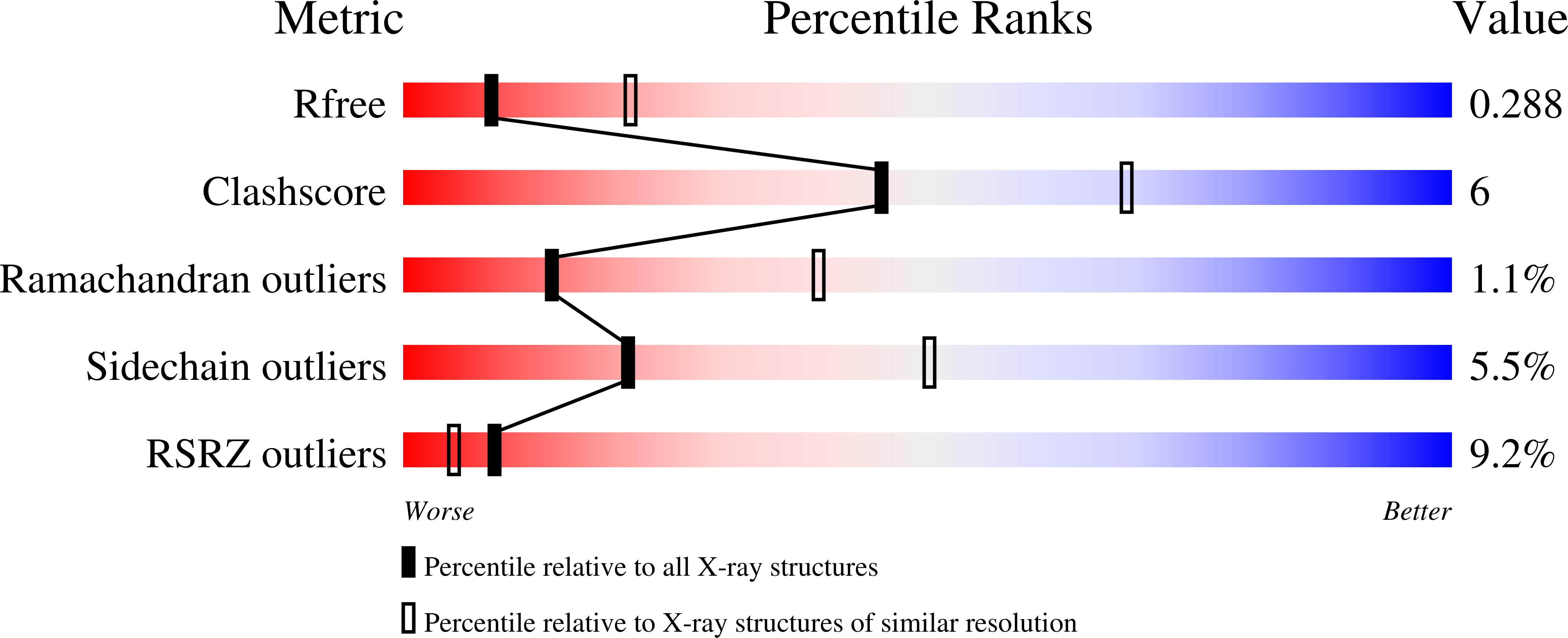
Resolution:
2.81 Å
R-Value Free:
0.28
R-Value Work:
0.23
R-Value Observed:
0.23
Space Group:
P 1 21 1
Macromolecular Entities
Structures with similar UniProt ID
Protein Blast
Polymer Type:polypeptide(L)
Molecule:Aspartic protease Bla g 2
Mutagens:N98Q
Chain IDs:A, B
Chain Length:330
Number of Molecules:2
Biological Source:Blattella germanica
Polymer Type:polypeptide(L)
Molecule:Antibody light chain
Chain IDs:C, E
Chain Length:211
Number of Molecules:2
Biological Source:Mus musculus
Polymer Type:polypeptide(L)
Molecule:Antibody heavy chain
Chain IDs:D, F
Chain Length:213
Number of Molecules:2
Biological Source:Mus musculus
Modified Residue
Compound ID Chain ID Parent Comp ID Details 2D Image
ASN A ASN GLYCOSYLATION SITE
CSX A CYS S-OXY CYSTEINE
Primary Citation
Crystal structure of a dimerized cockroach allergen Bla g 2 complexed with a monoclonal antibody.
J.Biol.Chem. 283 22806 22814 (2008)
PMID: 18519566 DOI: 10.1074/jbc.M800937200

Abstact

The crystal structure of a 1:1 complex between the German cockroach allergen Bla g 2 and the Fab' fragment of a monoclonal antibody 7C11 was solved at 2.8-angstroms resolution. Bla g 2 binds to the antibody through four loops that include residues 60-70, 83-86, 98-100, and 129-132. Cation-pi interactions exist between Lys-65, Arg-83, and Lys-132 in Bla g 2 and several tyrosines in 7C11. In the complex with Fab', Bla g 2 forms a dimer, which is stabilized by a quasi-four-helix bundle comprised of an alpha-helix and a helical turn from each allergen monomer, exhibiting a novel dimerization mode for an aspartic protease. A disulfide bridge between C51a and C113, unique to the aspartic protease family, connects the two helical elements within each Bla g 2 monomer, thus facilitating formation of the bundle. Mutation of these cysteines, as well as the residues Asn-52, Gln-110, and Ile-114, involved in hydrophobic interactions within the bundle, resulted in a protein that did not dimerize. The mutant proteins induced less beta-hexosaminidase release from mast cells than the wild-type Bla g 2, suggesting a functional role of dimerization in allergenicity. Because 7C11 shares a binding epitope with IgE, the information gained by analysis of the crystal structure of its complex provided guidance for site-directed mutagenesis of the allergen epitope. We have now identified key residues involved in IgE antibody binding; this information will be useful for the design of vaccines for immunotherapy.

Legend

Protein

Chemical

Disease

Primary Citation of related structures
Feedback Form
Name
Email
Institute
Feedback