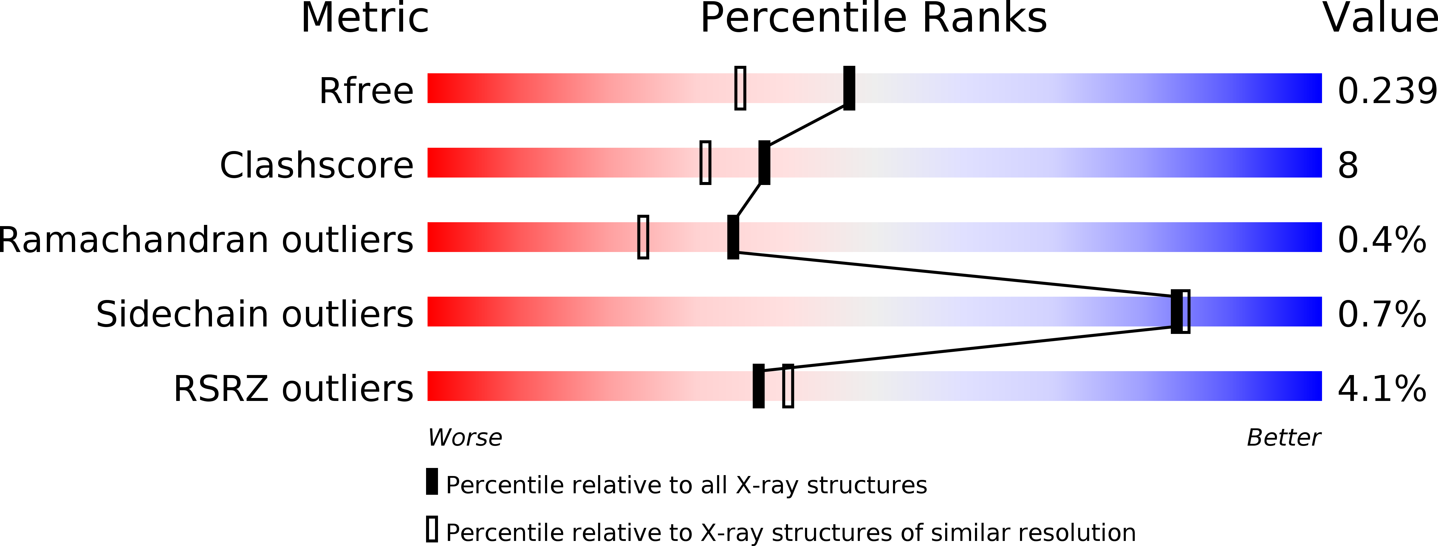
2NQO image
Deposition Date 2006-10-31
Release Date 2006-11-21
Last Version Date 2023-08-30
Entry Detail
PDB ID:
2NQO
Keywords:
Title:
Crystal Structure of Helicobacter pylori gamma-Glutamyltranspeptidase
Biological Source:
Source Organism:
Host Organism:
Method Details:
Experimental Method:
Resolution:
1.90 Å
R-Value Free:
0.23
R-Value Work:
0.19
Space Group:
P 1 21 1
Macromolecular Entities
Polymer Type:polypeptide(L)
Molecule:Gamma-glutamyltranspeptidase
Gene (Uniprot):HP_1118
Mutations:Engineered N-terminal histidine tag
Chain IDs:A, C
Chain Length:376
Number of Molecules:2
Biological Source:Helicobacter pylori
Polymer Type:polypeptide(L)
Molecule:Gamma-glutamyltranspeptidase
Gene (Uniprot):HP_1118
Chain IDs:B, D
Chain Length:188
Number of Molecules:2
Biological Source:Helicobacter pylori
Primary Citation
Autoprocessing of Helicobacter pylori gamma-glutamyltranspeptidase leads to the formation of a threonine-threonine catalytic dyad.
J.Biol.Chem. 282 534 541 (2007)
PMID: 17107958 DOI: 10.1074/jbc.M607694200

Abstact

Helicobacter pylorigamma-glutamyltranspeptidase (HpGT) is a glutathione-degrading enzyme that has been shown to be a virulence factor in infection. It is expressed as a 60-kDa inactive precursor that must undergo autocatalytic processing to generate a 40-kDa/20-kDa heterodimer with full gamma-glutamyl amide bond hydrolase activity. The new N terminus of the processed enzyme, Thr-380, is the catalytic nucleophile in both the autoprocessing and enzymatic reactions, indicating that HpGT is a member of the N-terminal nucleophile hydrolase superfamily. To further investigate activation as a result of autoprocessing, the structure of HpGT has been determined to a resolution of 1.9 A. The refined model contains two 40-kDa/20-kDa heterodimers in the asymmetric unit and has structural features comparable with other N-terminal nucleophile hydrolases. Autoprocessing of HpGT leads to a large conformational change, with the loop preceding the catalytic Thr-380 moving >35 A, thus relieving steric constraints that likely limit substrate binding. In addition, cleavage of the proenzyme results in the formation of a threonine-threonine dyad comprised of Thr-380 and a second conserved threonine residue, Thr-398. The hydroxyl group of Thr-398 is located equidistant from the alpha-amino group and hydroxyl side chain of Thr-380. Mutation of Thr-398 to an alanine results in an enzyme that is fully capable of autoprocessing but is devoid of enzymatic activity. Substrate docking studies in combination with homology modeling studies of the human homologue reveal additional mechanistic details of enzyme maturation and activation, substrate recognition, and catalysis.

Legend

Protein

Chemical

Disease

Primary Citation of related structures