Deposition Date
2006-10-31
Release Date
2007-07-24
Last Version Date
2024-12-25
Entry Detail
PDB ID:
2NQD
Keywords:
Title:
Crystal structure of cysteine protease inhibitor, chagasin, in complex with human cathepsin L
Biological Source:
Source Organism(s):
Trypanosoma cruzi (Taxon ID: 5693)
Homo sapiens (Taxon ID: 9606)
Homo sapiens (Taxon ID: 9606)
Expression System(s):
Method Details:
Experimental Method:
Resolution:
1.75 Å
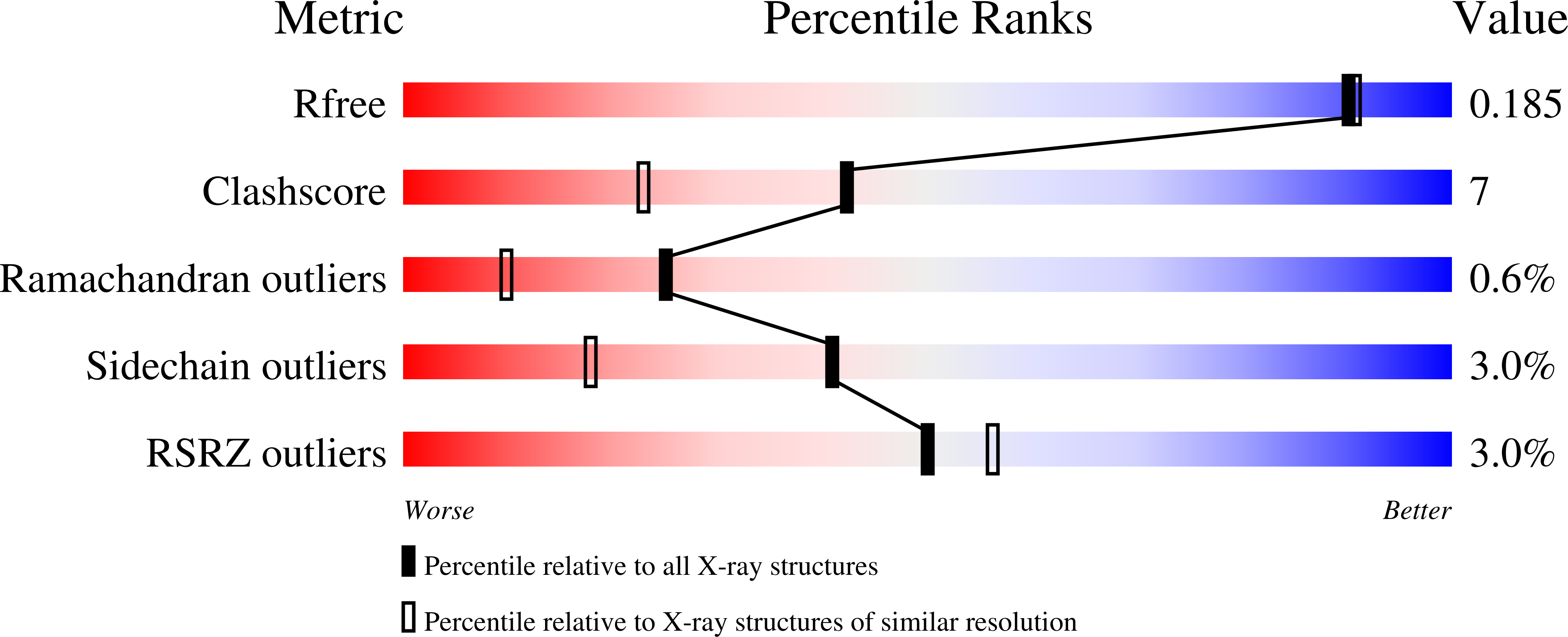
R-Value Free:
0.18
R-Value Work:
0.14
R-Value Observed:
0.15
Space Group:
P 1 21 1