Deposition Date
2006-10-26
Release Date
2006-12-26
Last Version Date
2024-10-16
Entry Detail
PDB ID:
2NP0
Keywords:
Title:
Crystal structure of the Botulinum neurotoxin type B complexed with synaptotagamin-II ectodomain
Biological Source:
Source Organism(s):
Mus musculus (Taxon ID: 10090)
Clostridium botulinum (Taxon ID: 1491)
Clostridium botulinum (Taxon ID: 1491)
Method Details:
Experimental Method:
Resolution:
2.62 Å
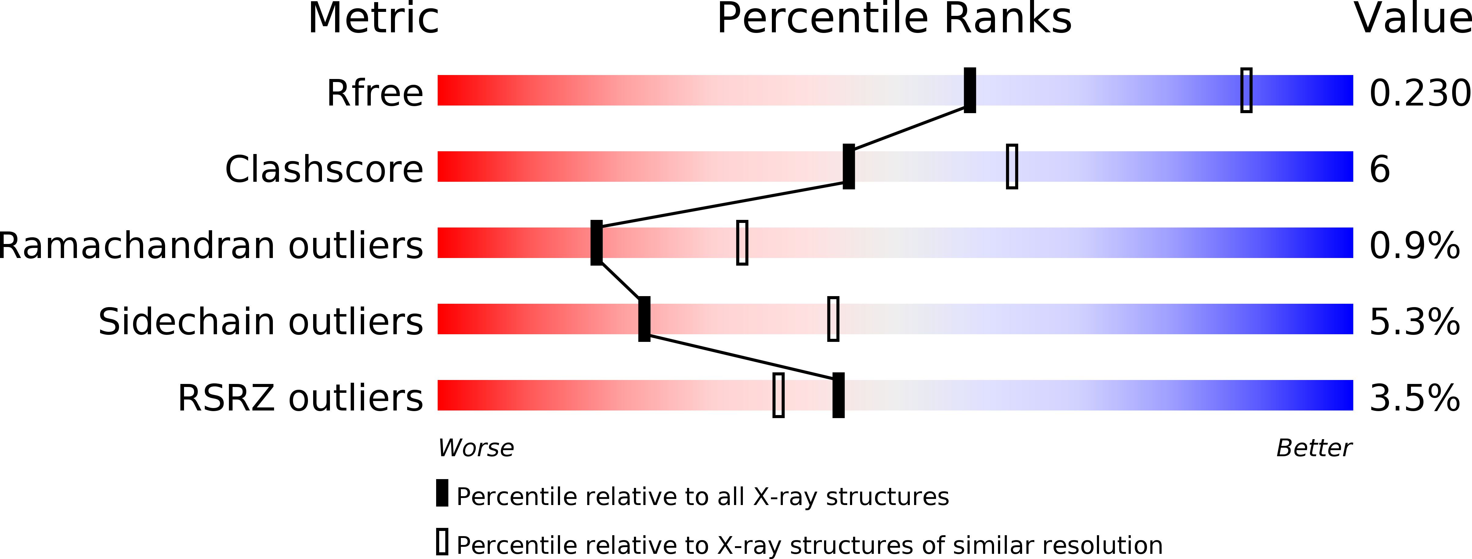
R-Value Free:
0.22
R-Value Work:
0.18
R-Value Observed:
0.18
Space Group:
C 2 2 21