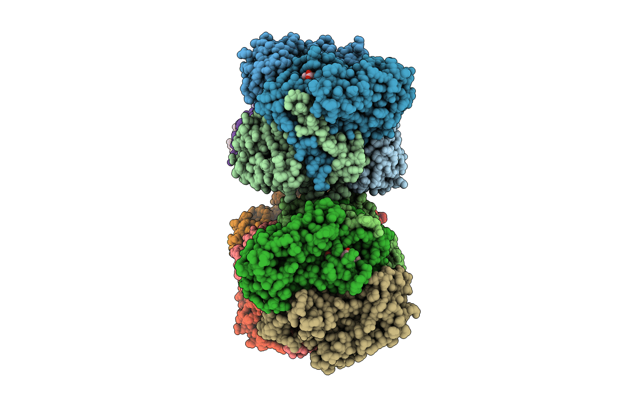
2NOX image
Deposition Date 2006-10-26
Release Date 2006-12-19
Last Version Date 2023-08-30
Entry Detail
PDB ID:
2NOX
Keywords:
Title:
Crystal structure of tryptophan 2,3-dioxygenase from Ralstonia metallidurans
Biological Source:
Source Organism:
Method Details:
Experimental Method:
Resolution:
2.40 Å
R-Value Free:
0.27
R-Value Work:
0.21
R-Value Observed:
0.21
Space Group:
P 1
Macromolecular Entities
Polymer Type:polypeptide(L)
Molecule:Tryptophan 2,3-dioxygenase
Gene (Uniprot):kynA
Chain IDs:A, B, C, D, E, F, G, H, I, J, K, L, M, N, O, P
Chain Length:281
Number of Molecules:16
Biological Source:Cupriavidus metallidurans
Ligand Molecules
Primary Citation
Crystal structure and mechanism of tryptophan 2,3-dioxygenase, a heme enzyme involved in tryptophan catabolism and in quinolinate biosynthesis.
Biochemistry 46 145 155 (2007)
PMID: 17198384 DOI: 10.1021/bi0620095

Abstact

The structure of tryptophan 2,3-dioxygenase (TDO) from Ralstonia metallidurans was determined at 2.4 A. TDO catalyzes the irreversible oxidation of l-tryptophan to N-formyl kynurenine, which is the initial step in tryptophan catabolism. TDO is a heme-containing enzyme and is highly specific for its substrate l-tryptophan. The structure is a tetramer with a heme cofactor bound at each active site. The monomeric fold, as well as the heme binding site, is similar to that of the large domain of indoleamine 2,3-dioxygenase, an enzyme that catalyzes the same reaction except with a broader substrate tolerance. Modeling of the putative (S)-tryptophan hydroperoxide intermediate into the active site, as well as substrate analogue and mutagenesis studies, are consistent with a Criegee mechanism for the reaction.

Legend

Protein

Chemical

Disease

Primary Citation of related structures